Tuesday 31 August 2021

Role of Temperature and Humidity in the Modulation of the Doubling Time of Covid-19 Cases

Role of Temperature and Humidity in the Modulation of the Doubling Time of Covid-19 Cases by Oliveiros B* in Open Access Journal of Biogeneric Science and Research


Abstract

  • Objectives: To estimate influence of meteorological variables, notabl temperature and relative humidity, on the spreading of the SARS- CoV-2 virus. Adjusted odds ratio were used to quantify the influence of temperature and relative humidity on the doubling time of the spread.
  • Setting: The data regarding the China COVID-19 outbreak used in this work were retrieved from the Johns Hopkins University COVID-19 dashboard. The meteorological data was obtained using the Meteostat Application Programming Interface.
  • Participants: No patients were directly included in this study since data were collected from internet sites. Primary and secondary outcome measures: the primary outcome measured was the number of confirmed infected and the secondary outcomes were temperature, relative humidity, wind speed and precipitation of the China provinces in the period of analysis.
  • Results: Results indicate that the doubling time correlates positively with temperature and inversely with relative humidity. We found that, at maximum, an average temperature increase of 1ºC augments the doubling time in 0.090 days and an increase of 1% of relative humidity leads to a decrease of 0.072 days in the doubling time.
  • Conclusion: The results obtained suggest that decrease in the rate of progression of COVID-19 with the arrival of spring and summer in the north hemisphere since a 20ºC increase is expected to delay the doubling time in 1.8 days in average. Temperature and relative humidity only explain 18% of the variation in disease doubling time; the remaining 82% may be related to containment measures, general health policies, population density, transportation or cultural aspects.
  • Trial Registration: Not applicable.

Strengths of this Study

  1. It is established a quantitative relationship between the rate of progression of COVID-19 and temperature and relative humidity
  2. The model is easily generalized to other situations

Limitations of this Study

    1. Only the exponential portion of the outbreak is used to determine the relationship between the variables
    2. There is uncertainty linked to the meteorological variables concerningtheir regional representatively as only one value is used to characterized a whole region

Keywords: COVID-19; doubling time; temperature; relative humidity; precipitation; wind speed; mortality

Introduction

The outbreak of pneumonia cases in Wuhan, China during last December led to great efforts to prevent a global epidemic. The alert from China CDC was rapidly transmitted to the World Health Organization [1-3] excluding possible causes such as influenza, adenovirus, severe acute respiratory syndrome (SARS-CoV) and Middle-East respiratory syndrome (MERS-CoV)[1,3-4]. The novel coronavirus, named SARS-CoV-2, and its genomic characterization was performed a few days after, permitting to devise a robust test method [1-5]. Although the genomic characterization revealed some relations both to SARS- CoV and MERS-CoV, [4-6] the new virus was found to be much more aggressive than those other coronaviruses or the seasonal one [4-6]. When human-to-human transmission was proved, on the 20th of January, the onset of the disease (COVID-19) has changed [2,7-10]. According to the China CDC11, the case fatality rate (CFR) was 0.2% at the end of January 2020 and 14.4% of the confirmed cases were considered severe or even critical. In the last week of February,79441 cases were confirmed worldwide (97% in Mainland China) and the number of deaths was 2620 (95% in Mainland China12,13). The epidemiological curves of COVID-19 in China showed the progression of illness in the outbreak over time from December 8, 2019 up to February 11, 2020,11 when there was a total of 72314 confirmed cases as the geo-temporal spread of COVID-19 [11].

At that time, the majority of confirmed cases occurred in the norther hemisphere and until the last week of February 2020 no confirmed cases had been reported in South America or Africa, except for one case in Egypt [12,13]. In fact, the first confirmed case in Brazil was reported on February 26, while Algeria and Nigeria reported the first cases respectively on the 25th and 27th of February. The discussion about the COVID-19 epidemic spread in the northern hemisphere, while low temperatures and high relative humidity are present, and the unknown, although expectable positive impact of spring and summer in sustaining             the epidemic, as its spread into the southern hemisphere was not as fast, has raised our question: how do meteorological variables, such as temperature and relative humidity, modulate COVID-19 duplication time?

Even though there is not yet strong evidence that meteorological conditions may have a role on COVID-19 outbreak or on human transmission, some studies have reported their role in guinea pigs’ influenza transmission [14] and enveloped virus survival [15] in droplets. Some evidence of a faster spreading of diseases in high relative humidity levels has been reported [16], namely for the Legionella disease, although this infection is not caused by a virus. Some papers have been written since mid-February on this topic, [17-20] even though the relationship is not perfectly established and more research is required. We intend to add value to this discussion by evaluating the meteorological impact on COVID-19 duplication time.

Material and Methods

Patient and Public Involvement

No patient involved

Methods              

The statistical model developed was implemented in two steps: firstly, an exponential model relating the accumulated number of confirmed cases and time was considered. Secondly, the rate of spread was used as dependent variable in a linear model that took as independent variables temperature, relative humidity, precipitation and wind speed. Only cases belonging to China were considered, as an attempt to control both cultural aspects and policies adopted to contain the virus. Therefore, data from the 31 provinces of Mainland China were gathered from the 23rd January up to the 1st of March, completing 39 days. These data were completed with meteorological variables, comprising temperature, relative humidity, precipitation and wind speed, collected for the same period, using the Meteostat Application Programming Interface (API) [21]. We searched for meteorological stations containing hourly measurements of these variables for the whole 39-day time period that were closest to the latitude and longitude coordinates that were available in the files that contained the confirmed cases time-series [13]. These geolocalization coordinates correspond to the geometrical centerpoint of each chinese province [22]. When meteorological data from a station was not possible to obtain around that position, another search point was chosen randomly from the set of closest nodes of an XY grid of nodes separated by 0.5 degrees in latitude and longitude and centered in the originally desired geolocation.

To compute the rate of spread a simple exponential model was assumed,

described by:

=,             (eq1)

0

where 0 is the number of infected at instant zero, represents the rate of infection or the rate of spread and is the time. A more natural way of interpreting is by transforming it into the doubling time, , given by:

=             (2)         .               (eq2)

The doubling time is the time needed to duplicate the number of infected subjects. Since the rate of progression changes over time and the exponential modelndoes not hold any longer, we considered mainly the initial days of the time series, selecting several periods of time     composed each one of a predetermined number of consecutive days, but with different starting points. The starting point was assumed by randomly choosing the first day of the period. For each province several periods were sampled, allowing to obtain more than one value of the rate of progression (Figure 1).

Two different periods of time of 12 and 15 days were tested, along with 3 and 155 different starting points.

For each rate of progression, a corresponding value of each meteorological variable was computed, taking into account the same period of time. We opted to use the median of the meteorological variables (temperature, relative humidity, precipitation and wind speed) as it is more robust than the mean and tends to better represent the central tendency of the variable. Only models attaining more than 0.75 for the adjusted R square value were selected (Figure 1).

The rate of progression was transformed into the doubling time, and recorded along with the median of temperature, relative humidity, precipitation and wind speed. These values were then used to fit a linear regression model aiming to assess how the meteorology is related to the doubling time. The exponential models for the rate of progression were computed resorting to the R programming language [23], whereas the linear regression models were computed using IBM SPSS v25 with an adopted statistical significance level of

0.05.

Figure 1: Flowchart of the routine in R language used to Compute the statistical models of Covid -19 Spreading of new cases.
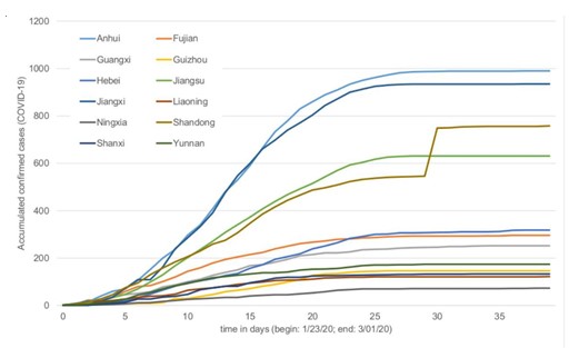
Figure 2: Accumulated confirmed cases of Covid-19 in function of time. Only the Provinces that had one or two cases at the beginning at the series are shown.

Figure 3: Doubling time for each province considering the different forms of calculation.
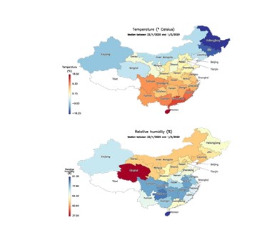
Figure 4:  Median temperature (top) and relative humidity (bottom) of each province during the studied period. The corresponding values are shown in Annex I.
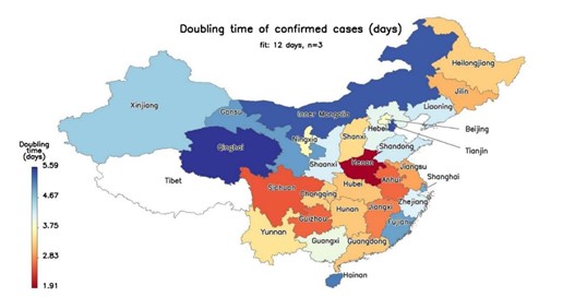
Figure 5: Doubling times obtained with one of the models (12 days, n=3).

Table 1: Statistics of the doubling time for two different time periods and two repetitions.

Table 2: Results of the linear regressions between doubling time, temperature and relative Humidity.

Results

The number of confirmed infected cases of COVID-19 were initially analysed by plotting them against time. (Figure 2) depicts the curves obtained for the provinces that at the first time point (23td of January) had only one or two cases. The analysis of (Figure 2) shows that the number of accumulated cases is different between provinces and, in general, its rate decreases over time up to the point where it becomes null. Since the objective of the study was to analyse the rate of spread, we decided to use periods of 12 and 15 days to determine it. The initial 25 days were thought to be the most informative regarding the rate of spread. The fits of temporal evolution of confirmed cases of COVID-19 of the remaining provinces are not shown, but a similar profile can be obtained, leading to the same conclusion.

A two way ANOVA shows statistical differences (F(1, 450)=23.573; p<0.001) between the size, 12 or 15 days of the period employed to compute the doubling time, but no statistical differences (F(1, 450)=0.047; p=0.828) between the number of samples taken from each province. This result is in agreement with the hypothesis that the doubling time changes with time. The average of the doubling time duplication was determined for each province (Figure 3).

Taking the values of doubling time and the meteorological variables, a linear regression was performed. (Table 2) shows, for the 4 conditions studied, the results achieved from the linear regressions, that assumed temperature and relative humidity as independent variables. Precipitation and wind speed did not reach statistical significance in any model (data not shown), thus (Table 2) only refers results for temperature and relative humidity.

The results obtained for all models are statistically meaningful and despite their variation, it is possible to perceive that the coefficients of regression (B) are not statistically different as their confidence intervals overlap. On the other hand, the amount of variation explained, given by the adjusted R square value, differs between models. The model based on 12 days and 3 sampled periods is able to explain 18% of the variance in the doubling time, which means that temperature and relative humidity alone may describe 18% of the variation of confirmed COVID-19 infections. More importantly, the signal and value of the coefficients of regressors are of utterly importance to understand how the spread of COVID-19 is expected to be affected by temperature and relative humidity. According to all models, temperature increases the doubling time, which means that it delays the spread of COVID-19. Relative humidity, on the contrary, benefits it. The models differ, however, on the amount of contribution: for example, in the best scenario (model: 15 days, n=3) the doubling time is increased by 0.090 days for each Celsius degree increase, and is increased by 0.072 days for each unit decrease of the relative humidity value (Figures 4 & 5).

Discussion and Conclusion

In this work, the way temperature and relative humidity affect the doubling time of COVID-19 spreading was determined. Results suggest that temperature correlates positively with the doubling time and negatively with relative humidity. This means that, with spring and summer, the rate of progression of COVID-19 is expected to be slower. Still, these two variables contribute at maximum to 18% of the variation, being the remaining 82% related to other factors such as containment measures, general health policies, population density, transportation, cultural aspects, etc. Besides, the direct impact is also small: for example, if temperature raises 20ºC, it is expected that in the best-case scenario the doubling time increases on average

1.8 days. These results are in agreement with other studies that suggested that the aerosol spread of the influenza virus is both dependent upon relative humidity and temperature, although performed in animal models [14], and that the virus survival in droplets is higher at high relative humidity levels with a significant

decrease on its infectivity rate at mid-levels of relative humidity [15].

Wei Lo et al 17 recently reported a statistically significant association between absolute relative humidity and mean temperature on COVID-19 spread among China provinces. Furthermore, they have concluded that transmission and exponential growth of confirmed cases are occurring in China provinces in relative humidity conditions ranging from cold and dry (Jilin or Heilongjiang) to tropical (Guangxi or Singapore), suggesting that changes in weather, as expected by the arrival of spring and summer, will not necessarily lead to declines in outbreak unless extensive public health interventions are implemented, and that further studies on the effects of meteorology on COVID-19 transmission are needed.

On the other hand, Jin Bu et al 18 reached the conclusion that continuous warm and dry weather is conducive to the survival of the 2019-nCoV and speculate that conditions such as temperature ranging from 13 to 19°C and relative humidity between 50% and 80% are suitable for the survival and transmission of this new coronavirus. However, their predictions were performed using SARS data and meteorological conditions at that time and, as they report, 2019-nCoV has a high basic case reproduction number (R0) lying between 2.2 to 6.7, causing much more infections than SARS.

Moreover, Mao Wang et al 19 have recently submitted a paper supporting that temperature could change the COVID-19 transmission and that there might be an optimal temperature for the viral transmission, suggesting that colder regions in the world should adopt strictest control measures. Yuwen Cai [20] did not find any correlation between the growth rate of the epidemy and daily mean temperature in either Wuhan or Hunan but found a weak correlation between the mortality in confirmed cases and daily mean temperature both in Wuhan (r = -0.441) and Hubei (r = -0.440), although not adjusted for the useof three makeshift hospitals, which proved to be effective. The main focus of this work was to assess the relationship between the rate of spread of COVID-19 and some meteorological variables, which determines the type of model adopted.

Although the reproduction number, 0, is the parameter widely accepted to characterize the velocity of spreading, there are different forms of computing it, which tend to lead to different results [24]. On the other hand, the 0 calculation is generally based on assumptions about the epidemic phenomenon such as serial interval distribution [25] or“ the population is closed, that all cases are observed, and use daily case counts only” [26]. For the reasons mentioned, we opted for a simple/naive model that could assimilate the principal aspects of the variation of COVID-19 cases and translate it into a straightforward measurement that could be easily comprehended. Obviously, this model has several  drawbacks, mostly regarding the optimal period where an exponential growth is verified. We studied two different periods sizes and a random starting point aiming to analyse the impact of this aspect and        as an attempt to  mitigate its consequences. The doubling time values vary with the period size 26% at maximum (Table 1), which is unneglectable. Even so, this difference only affects slightly the regression coefficients of temperature and relative humidity, since they do not show statistical differences. The average incubation time accepted for the COVID-19 is 3 days, but it can be as long as 14 days. This is a problem, since when the results of the tests are provided, the transmission has already occurred and the meteorological variables should be given for that period. Therefore, the use of a random starting point and two periods to compute the duplication time are a form to compensate the uncertainty caused by the incubation time. This effect is particularly important for the first points in each period, which is balanced by the use of different starting points, and a time period of 15 days that can accommodate the great majority of transmission. On the other hand, as we are looking at the first days of the epidemic with exponential growth, the later points have more weight than the initial ones. Moreover, we use the median of temperature and relative humidity, instead of the average, of this random sliding time window because it is a robust measure against outliers and translates 50% of the results. Besides, the maximum interquartile range that we found was 4.4ºC for temperature and 29.1% for relative humidity (Annex I).

Another point of possible bias is the COVID-19 data that do not cover all provinces from the beginning of the outbreak, making it difficult to homogeneously study in all provinces the time period that corresponds to the exponential growth. A different concern is the possibility that containment measures may affect the results, and that was the reason why we selected China. To obtain meteorological variety we needed a vast region that could be the concatenation of several countries or just one large country. China seems to be the most appropriate since it has a vast territory that is governed under the same main rules and that adopted similar containment measures and testing policies in a short period of time. Regional differences certainly exist and this could be one of the factors that the linear models did not capture, as we         mentioned before. Nonetheless, it is accepted that the results of containment measures have a delay of at least 2 weeks, and since we are analyzing the first days, these effects would not interfere with our analysis.

The meteorological variables in this study were obtained for locations near the center of provinces, which typically do not correspond to the average location of the population. Measurements that better represent the central tendency of the meteorological variables felt by the general population of a particular region are currently being implemented. A final remark about the short average doubling time values obtained (3.78 to 4.53 days - Table 1), which should be a motive of concern. For each doubling time, the number of infected doubles, so one month of sustained growth at a conservative pace of 5 days means an increase of the number of infected by a factor of 64.

Contributor Ship Statement

All the    authors have equally contributed in the implementation       of the computational model, assessment of the results and writing of the manuscript.

Competing Interests

There are no competing interests.

Funding

Funded by National Funds via FCT (Foundation for Science and Technology) through the Strategic Project                UIDB/04539/2020 and    UIDP/04539/2020 (CIBB).

Data Sharing Statement

Data used in this work are publicly available and cited accordingly.

More information regarding this Article visit: OAJBGSR


Tuesday 24 August 2021

Anesthesia in Obesity

Anesthesia in Obesity by Özgür Oğul Koca in Open Access Journal of Biogeneric Science and Research


Review Article

Ideal weight (kg) = Height (cm) -100 (Male)

Ideal weight (kg) = Height (cm) -105 (Female)

Obesity 20% from ideal weight ↑

In TURKEY 35% of total population obese, 6% morbidly obese 38.5% of women 26.4% of men are obese

Obese (BMI 30 - 34.9)

Severely Obese (BMI 35 -39.9)

Morbid Obese (BMI> 40)

Super Obese (BMI> 50)

Ultra Obese (BMI> 60)

Mega Obese (BMI> 70)

  1. Obesity-related problems
  2. Pulmonary embolism
  3. DVT
  4. Cancer
  5. Stroke
  6. Sleep Apnea Syndrome
  7. Osteoarthritis
  8. Type 2 DM
  9. Hypertension
  10. Coronary artery disease
  11. Metabolic syndrome
  12. Hypoventilation syndrome
  13. Airway and respiratory system affected [1]
  14. Reproductive problems
  15. Liver and gall bladder diseases

Increased cardiac output and blood volume cause an increase in the workload of the heart. Arterial hypertension and left ventricular hypertrophy develop because the increase in cardiac output (0.1 L / min for every 1 kg of adipose tissue) is provided by the increase in stroke volume. It causes pulmonary vasoconstriction due to increased pulmonary blood flow and hypoxia, pulmonary hypertension and corpulmonary.

Obesity-hypoventilation Syndrome (Pickwickian syndrome) is a complication of extreme obesity. It is characterized by hypercapnia, polycythemia due to hypoxia, right heart failure and sleepiness. These patients have weak respiratory stimulation and snoring and upper airway obstruction (obstructive sleep apnea syndrome) are observed during sleep. Obstructive sleep apnea syndrome causes an increase in perioperative complications: hypertension, hypoxia, dysrhythmia, MI, pulmonary edema, difficulty in airway patency during induction, airway obstruction during recovery If opioids and sedatives are used and the supine position is given, the risk of postop airway obstruction is high. Therefore, CPAP application should be considered until full recovery is achieved [1,2].

Factors Affecting Drug Distribution in Obesity

Increased cardiac output, increased blood volume, increased organ size and increased fat mass. Theoretically, excess fat deposits cause an increase in the distribution volume of fat-soluble drugs (benzodiazepines, opioids, thiopental, propofol). The increase in volume of distribution means that a higher loading dose is required for the same plasma concentration. However, the restricted blood flow of adipose tissue reduces the effects of increased adipose tissue on acute distraction and elimination of the drug.^[3] The distribution volumes and elimination half-lives of lipophilic drugs increased in obese patients.

The response of the central nervous system to the induction dose of thiopental in obese patients is not different from that of non-obese patients, so the dose of the drug should be chosen according to the ideal body weight, not the actual weight of the patient.

Summary

The dose of intravenous induction agents should be adjusted according to the needs of the patient, not by calculation of milligrams per kilogram. Since the clearance rate is expected to decrease due to the high volume of distribution, the frequency of maintenance drug administration should also be reduced. The distribution volume of the water-soluble drugs (neuromuscular blockers) did not change. However, to avoid drug overdose, water-soluble drugs should be given according to ideal body weight. The distribution of volatile anesthetics to adipose tissue is very slow. Volatile anesthetics can be stored in adipose tissue. However, prolongation of recovery is not expected from volatile anesthesia in obese patients due to the slow distribution to adipose tissue. Increased metabolism of volatile agents and hypoxia in obese patients explain the increased risk of halothane hepatitis in these cases [4]

  1. Isoflurane and desflurane can be chosen in obese cases as they are the least metabolized volatile agents.
  2. Caution should be exercised in using nitrous oxide in obese cases due to increased intrapulmonary shunts and oxygen requirement.
  3. Care should be taken in the use of opioids due to the increased risk of postoperative hypoxemia and hypoventilation.
  4. Story
  5. Sleep apnea
  6. Somnolence
  7. HT, CHF, coronary artery diseases
  8. GER, hiatal hernia
  9. DM
  10. Deep vein thrombosis
  11. Physical examination
  12. Respiratory system: Dyspnoea, orthopnea, cyanosis
  13. Airway should be evaluated; Sits and is in a supine position
  14. The neck is short and thick
  15. Temporomandibular and atlantooccipital joint movements are limited
  16. The top airlines are narrow
  17. The distance between the mandible and the sternal fat pads is short
  18. Pharyngeal and palatal soft tissues are abundant
  19. Larynx may be in anterior localization
  20. Language is big

Cardiovascular System

Hypertension, heart failure, angina It should be evaluated in terms of arterial and vein access. Large blood pressure cuff (cuff should cover 70% of the arm)

Arterial Catheter Tests

ECG Ac radiography Detailed biochemistry (KC func, Lipid, blood sugar etc.)

Blood Gases Respiratory Function Tests Position

20-30 Reverse Trendelenburg: Ideal Premedication: Gastric acidity (H2 antagonists, anticides) and gastric volume (metoclopramide) should be reduced Sedatives, hypnotics and opioids should be used with caution due to sleep apnea. Intubation [1,3]

Awake endotracheal intubation may be safe in patients with massive obesity, small mouth-short neck, sleep apnea, and patients with impaired pulmonary and cardiovascular function.

Fiberoptic intubation may be required.The ramp position can facilitate intubation. In obese patients, desaturation may develop rapidly during the apnea period during intubation, as lung volumes are decreased and oxygen consumption is increased.Therefore, the cases should be preoxygenated before induction and denitrogenation of the lungs should be provided. Induction agents should be short acting. Intubation should be confirmed with end-tidal carbon dioxide, as respiratory sounds may not be heard well.

Ventilation [1,2,3].

General anesthesia can worsen oxygenation by causing a decrease in functional residual capacity and impairment in the ventilation-perfusion relationship. Therefore, controlled ventilation with 50% oxygen is frequently applied in these cases. In these cases, controlled ventilation with high tidal volume provides better oxygenation. Even with lithotomy, trendelenburg and controlled ventilation in the prone position, sufficient oxygenation may not be achieved and the oxygen concentration is increased in these cases. PEEP should be used with caution. Excessive levels of PEEP may further increase existing pulmonary hypertension.

Regional Anesthesia

Due to the adipose tissue, the cue points are unclear so there may be a hassle In obese cases, the dose of local anesthetic to be used for epidural and spinal anesthesia should be 20-25% less than normal individuals, since epidural adipose tissue is excessive and epidural veins are large. In the sitting position, the localization of the midline and the insertion of the spinal needle is easier. Postop respiratory complications are less in regional anesthesia.

Postop Prefer regional techniques for pain control. Patient controlled analgesia may also be preferred. Be wary of respiratory depression. Make sure that the muscle relaxant effect is fully antagonized (perform neuromuscular monitoring if necessary) Monitor oxygenation with a pulse oximeter Position in a half-seated (45 degrees) recovery room (diaphragm load is reduced) The risk of hypoxia may continue for a few days postoperatively; Oxygen should be given routinely. Early ambulation should be provided There are risks of postop wound infection, deep vein thrombosis and pulmonary embolism.

More information regarding this Article visit: OAJBGSR

Wednesday 18 August 2021

Mir-146a Inhibits IFN-Γ Production Via Suppressing TLR4/IRAK-1/NF-kb Expresion in Pulmonary Arterial Smooth Muscle Cells

Mir-146a Inhibits IFN-Γ Production Via Suppressing TLR4/IRAK-1/NF-kb Expresion in Pulmonary Arterial Smooth Muscle Cells by Shuyuan Chu in Open Access Journal of Biogeneric Science and Research


Abstract

Purpose: The microRNA-146a (miR-146a) could regulate proliferation of vascular smooth muscle cell and inhibits inflammation of airway, but its role in inflammation of pulmonary arterial smooth muscle cell (PASMC) hasn’t been reported. We aim to explore the effect of miR-146a on regulating inflammatory signaling in the study.

Methods: Primary PASMCs were separated from rats. Cells were stimulated by lipopolysaccharides (LPS). miR-146a was transfected into cells with plasmid. miR-146a expression in PASMCs was assessed by real-time PCR. The protein expression of TLR4, phosphorylated-IRAK-1, phosphorylated-IKK, phosphorylated-IκB and NF-κB (P65) in PASMCs was analyzed using western blotting. The level of IFN-γ was detected using ELISA.

Results: The protein expression of TLR4, phosphorylated-IRAK-1, phosphorylated-IKK, phosphorylated-IκB and NF-κB (P65) in PASMCs was increased when induced by LPS, which was reversed by miR-146a. The level of IFN-γ in supernatant of PASMCs was higher in LPS-treated group than controls, which was decreased in cells with miR-146a overexpressed.

Conclusion: miR-146a could attenuate LPS-induced IFN-γ production, and activation of TLR4, IRAK-1 and NF-κB in PASMCs, which might provide novel target on the therapy of pulmonary hypertension.

Introduction

Pulmonary hypertension (PH) is a hemodynamic and pathophysiologic syndrome from increased blood pressure within pulmonary arteries, which prevalence is approximately 10 % in general population. Its prognosis is depressed that the one-year mortality is approximately only 15% [1]. Pulmonary arterial smooth muscle cell (PASMC) participates in PH through activating inflammatory signaling, such as NF-κB pathway [2,3]. However, the precise mechanisms of inflammation in PASMC are not very clear.

Recent studies have showed that microRNA-146a (miR-146a) could regulate proliferation of vascular smooth muscle cell from aortic artery [4,5] In addition, miR-146a could reduce inflammation in airway by targeting on IRAK-1 [6]. However, the role of miR-146a in inflammation of PASMC hasn’t been reported. Interestingly, miR-146a was found to contribute to inhibiting lipopolysaccharides (LPS)-induced activation of TLR4/IRAK1/NF-κB signaling in monocytes [7]. Similar finding was showed in intestine epithelial cells [8]. These findings suggested that miR-146a could inhibit the activation of TLR4/IRAK1/NF-κB signaling in inflammation. Moreover, our previous work found that LPS could induce the activation of TLR4/IRAK-1/NF-κB signaling, resulting in an increased production of IFN-γ in PASMCs [9]. Thus, in this study, we explore the role of miR-146a in regulating IFN-γ production and TLR4/IRAK-1/NF-κB signaling activation in PASMCs.

Methods

Cell Culture and Transfection

Male Wistar rats (8-10 weeks old, weighing 280±20 g) were obtain from experimental animal center of Guilin Medical University. All experimental procedures were approved by the Animal Care and Use Committee of the Affiliated Hospital of Guilin Medical University. Rats were anaesthetized with 5% isoflurane by inhalation in oxygen and killed by cervical dislocation. The small vascular was separated from the 3rd level or lower artery branch of pulmonary lobe segments, and then was minced to small pieces and digested by 0.2% type I collagenase for 20 min at 37 °C in water. Digestion was stopped by adding 10% FBS (GIBCO, MA, USA)). The primary PASMCs were cultured in DMEM medium containing 10% FBS at 37°C in 5% CO2. Seven days later, PASMCs at passages 3-6 were used to conduct the experiments. Cells were cultured in serum-free medium 30min prior to transfection.

The primary PASMCs were identified using immunohistochemistry with α-SM-actin staining (Figure 1). The slides of cells were fixed by 4% paraformaldehyde for 20 min and incubated in 0.6% H2O2 for 30 min to quench endogenous peroxidase activity. The slides were incubated with primary mouse anti-rat antibody against α-SM-actin (dilution 1:100, BM0002, BOSTER, Wuhan, China) at 4 ◦C overnight, and then were incubated with horseradish peroxidase conjugated goat anti-mouse IgG antibody (BA1001, BOSTER, Wuhan, China) at room temperature for 20 min. After washes with PBS for three times, 3’3-diaminobenzidine-tetrahydrochloride was applied on the slides as a chromogen for 1–5 min, and were then by haematoxylin for 5–10 min. The transfection of miR-146a was performed with plasmid (Genechem, Shanghai, China) and lipofectamine2000 (Invitrogen, MA, US) according to the manufacturer’s instruction. When six hours after transfection, LPS-induced cells were stimulated with LPS(1μg/ml) (Sigma, MO, US) for 48 hours.

Quantitative Real-Time PCR

PASMCs (1×10⁶cells/well) were plated into six-well plates and incubated overnight in a humidified incubator at 37 ◦C in 5% CO2. Total RNA was extracted using the RNA simple Total RNA Kit according to the manufacturer’s protocol, and RNA was reverse transcribed into cDNA. A quantitative real-time polymerase chain reaction (PCR) was performed using Hairpin-itTM microRNA and U6 snRNA Normalization real time-PCR quantitation kit (GenePharma, Shanghai, China)) with ABI PRISM 7500 Sequense Detection System (Thermo Fisher Scientific, Inc, Carlsbad, California, USA) in accordance with the manufacturer's protocol. The 20μl PCR reactions (with 10μl Real-time PCR Master Mix, 0.4μl microRNA-146a /U6 snRNA specific Primer set, 0.2μl microRNA-146a /U6 snRNA specific Probe, 0.4μl ROX reference dye, 0.2μl Taq DNA polymerase, 2μl miRNA RT product and 6.8μl PCR H2O) were undergone 3 min at 95 ◦C, then 40 cycles of 12 s at 95 ◦C and 40 s at 62 ◦C. RT and PCR primer sequences are as follows: miR-146a RT: GTCGTATCCAGTGCGTGTCGTGGAGTCGGCAATTGCACTGGATACGACAACCCAT, miR-146a FP: GGCGTGAGAACTGAATTCCA, miR-146a RP: TCGTGGAGTCGGCAATTG; U6 RT: CGCTTCACGAATTTGCGTGTCAT, U6 FP: GCTTCGGCAGCACATATACTAAAAT, U6 RP: CGCTTCACGAA TTTGCGTGTCAT. The level of mRNA expression was reported as fold change using the 2–△△CT method. Every sample was triplicated.

Western Blot Analysis

PSMCs were treated with 100μl RIPA and PMSF (100:1) for 30 min on ice, and then centrifuged at 12000 × g (4 ◦C) for 20 min. The loaded proteins (15μg) were separated on a 10% SDS-polyacrylamide gel electrophoresis (SDS-PAGE), followed by transferring onto PVDF membranes. After blocking with 5% non-fat dried milk for 2 h at room temperature, the membranes were incubated with mouse anti-rat antibodies against TLR4 (dilution 1:500; Abcam, MA, USA)), IKK (dilution 1:5000; Abcam, MA, USA), β-actin (dilution 1:1000; ZSGB-Bio, Beijing, China), rabbit anti-rat antibodies against IRAK-1 (dilution 1:500; Abcam, MA, USA), IκB (dilution 1:1000; Abcam, MA, USA), and NF-κB (p65) (dilution 1:1000; CST, MA, USA) overnight at 4 ◦C, and then were incubated with horseradish peroxidase conjugated goat anti-mouse or anti-rabbit(dilution 1:5000) for 1 h. Finally, the blots were developed with the ECL Plus reagents (Bio-Rad, USA).

Enzyme-Linked Immunosorbent Assay

Enzyme-linked immunosorbent assay (ELISA) was used to detect the level of IFN-γ in cell culture supernatants according to the protocol of ELISA kit (Elabscience, Wuhan, China). Each sample was repeated in three wells. Briefly, in 96-well plates, 100μl sample and 100μl biotinylated detecting antibody (50μl cells and 50μl Detection reagent A) were incubated for 1 h at 37 °C, followed by incubation with 100μl Horseradish-peroxidase (HRP) conjugated working solution for 30 min at 37 °C. Subsequently, plates were incubated with substrate solution as a chromogen for 15 min without light. The optical density (OD) was measured at 450 nm using a microplate reader (TECAN, Switzerland).

Statistical Analysis

All statistical analyses were performed using SPSS 21.0 (IBM SPSS Inc., Chicago, IL, USA). Group data are expressed as mean ± std. deviation (SD). Significant differences were evaluated using an independent-samples t-test, and multiple groups were compared using one-way analysis of variance (ANOVA) followed by the Student–Newman–Keuls test or the Games–Howell test. p-values < 0.05were considered to be statistically significant.

Results

miR-146a Inhibits TLR4 Expression in PASMCs

When PASMCs were transfected with miR-146a, the expression of miR-146a was respectively increased about 6-fold at the 24th hour and 18-fold and at the 48th hour (Figure 1A). This demonstrated the successful transfection of miR-146a. Moreover, the expression of miR-146a was significantly induced by LPS after 24-hour administration (Figure 1B). That effect was time-dose dependent. Furthermore, the protein expression of TLR4 in PASMCs was detected after miR-146a transfection. TLR4 expression was increased in LPS group compared with controls, whereas it was reversed when transfected with miR-146a (Figure 2). Thus, miR-146a could inhibit TLR4 expression in PASMCs.

miR-146a Inhibits IRAK-1 Activation in PASMCs

The activation of IRAK-1 in PASMCs was detected by Western blotting. The protein expression of phosphorylated-IRAK-1 (Figure 3) was increased when treated with LPS. However, it’s reduced in cells with miRA-146a overexpression. These findings suggest that miR-146a could inhibit IRAK-1 activation in PASMCs.

miR-146a inhibits IKK activation in PASMCs

The activation of IKK, IκB and NF-κB (P65) in PASMCs was detected by western blotting. The protein expression of phosphorylated-IKK (Figure 4), phosphorylated-IκB (Figure 5) and NF-κB (P65) (Figure 6) was increased when treated with LPS. However, it’s reduced in cells with miRA-146a overexpression. These findings suggest that miR-146a could inhibit IKK activation in PASMCs.

miR-146a inhibits the secretion of IFN-γ in PASMCs

The level of IFN-γ in the supernatant of PASMCs culture medium was assessed by ELISA. (Figure 7) illustrates that the level of IFN-γ was higher in LPS group than controls. In contrast, it’s decreased in cells with miRA-146a overexpression. These findings indicate that miR-146 could inhibit IFN-γ secretion in PASMCs.

Figure 1: miR-146a expression in PASMCs. (A) miR-146a is overexpressed in PASMCs when transfected with Rno-mir-146a plasmids. The expression level of miR-146a was respectively increased about 6-fold at the 24th hour and 18-fold and at the 48th hour. (B) miR-146a expression is induced by LPS. The expression of miR-146a was significantly increased after 24 hours induced by LPS. That effect was time-dose dependent. *: p<0.05, **: p<0.01

Figure 2: miR-146a inhibits TLR4 expression in PASMCs. PASMCs were transfected miR-146a expressing plasmids. TLR4 expression in PASMCs was increased in LPS group, whereas it was reversed when transfected with miR-146a. The representative images are shown in left panel and quantitative analysis results are shown in right panel. **: p<0.01.

Figure 3: miR-146a inhibits IRAK-1 activation in PASMCs. PASMCs were transfected miR-146a expressing plasmids. Phosphorylated-IRAK-1 expression in PASMCs was increased in LPS group, whereas it was reversed when transfected with miR-146a. The representative images are shown in left panel and quantitative analysis results are shown in right panel. * p<0.05, ** p<0.01.

Figure 4: miR-146a inhibits IKK activation in PASMCs. PASMCs were transfected miR-146a expressing plasmids. Phosphorylated-IKK expression in PASMCs was increased in LPS group, whereas it was reversed when transfected with miR-146a. The representative images are shown in left panel and quantitative analysis results are shown in right panel. * p<0.05, ** p<0.01.

Figure 5: miR-146a inhibits IκB phosphorylation in PASMCs. PASMCs were transfected miR-146a expressing plasmids. Phosphorylated-IκB expression in PASMCs was increased in LPS group, whereas it was reversed when transfected with miR-146a. The representative images are shown in left panel and quantitative analysis results are shown in right panel. **: p<0.01.

Figure 6: miR-146a inhibits NF-κB (p65) expression in PASMCs. PASMCs were transfected miR-146a expressing plasmids.NF-κB (p65) expression in PASMCs was increased in LPS group, whereas it was reversed when transfected with miR-146a.The representative images are shown in left panel and quantitative analysis results are shown in right panel. **: p<0.01.

Figure 7: miR-146a inhibits the secretion of IFN-γ in PASMCs. PASMCs were transfected miR-146a expressing plasmids. Six hours after transfection, the cells were treated with LPS(1μg/ml) until 48 hours post-transfection. The cell culture medium supernatant was collected and IFN-γ production was measured by ELISA. The level of IFN-γ was increased when treated with LPS, which could be reversed in cells with miR-146a overexpression. *: P<0.05, **: P<0.01.

Discussions

Our study shows that miR-146a could attenuate LPS-induced IFN-γ production, TLR4 expression, and activation of IRAK-1 and NF-κB in PASMCs. The present study confirmed our previous finding that LPS could induce IFN-γ production, [9] and further found that miR-146a could significantly inhibit LPS-induced IFN-γ production. In vascular smooth muscle cells, IFN-γ could stimulate NF-κB activation, leading to inflammation [10] Those findings suggest that in vascular smooth muscle cells, IFN-γ may be not only an effector of LPS stimulation, but also a stimulator in the process of inflammation. It may play a key role in positive feedback of inflammation. Thus, it’s meaningful to disturb that feedback for reducing inflammation in PH treatment. The miR-146a may be a potential target since it could reduce LPS-induced IFN-γ production in PASMCs as it’s found in our study.

TLR4 is a crucial signaling in promoting inflammation of vascular smooth muscle cells [11-14]. Our study found that TLR4 expression in PASMCs was increased in LPS group, whereas it was reversed when transfected with miR-146a. The miR-146a could regulate TLRs and downstream signaling through TNF receptor-associated factor 6 and IL-1 receptor-associated kinase [15]. Thus, our findings suggest that miR-146a could suppress LPS-induced TLR4 expression in PASMCs.

Furthermore, TLR4 could activate NF-κB singling in LPS-induced inflammation of vascular smooth muscle cells from thoracic aortas [16,17]. Similarly, IRAK-1 is also the downstream of TLR4 in vascular smooth muscle cells from thoracic aortas [18]. In pulmonary vascular smooth muscle cells, TLR4 could activate IRAK-1/NF-κB signaling.9 Therefore, we explore the role of miR-146a in the activation of TLR4/IRAK-1/NF-κB signaling in PASMCs in the present study. This study showed that when miR-146a was overexpressed, the LPS-induced activation of IRAK-1 and NF-κB singling in PASMCs was inhibited. Since TLR4/IRAK-1/NF-κB singling in PASMCs could be activated by LPS and then lead to IFN-γ production, [9] we supposed that miR-146a could attenuate the activation of TLR4/IRAK-1/NF-κB singling, resulting in the decreased production of IFN-γ. Therefore, miR-146a may be a potential therapeutic target on inflammation of pulmonary artery, which may provide novel avenues in the therapy of PH.

Conclusion

In conclusion, miR-146a could attenuate LPS-induced IFN-γ production via inhibitingTLR4/IRAK-1/NF-κB pathway in pulmonary arterial smooth muscle cells, which might provide novel target on the therapy of PH.

More information regarding this Article visit: OAJBGSR

Sunday 15 August 2021

Prevalence and Associated Demographic Factors of Urinary Incontinence among Women Attending Postnatal Clinics in Primary Health Care In Qatar. A Descriptive Cross-Sectional Study

Prevalence and Associated Demographic Factors of Urinary Incontinence among Women Attending Postnatal Clinics in Primary Health Care In Qatar. A Descriptive Cross-Sectional Study by Shajitha Thekke Veettil* in Open Access Journal of Biogeneric Science and Research


Abstract

Background: Urinary Incontinence (UI) is more prevalent among women and is a significant health concern which affects the quality of life (QOL) of half of women of middle and older ages. The objectives of this study was to estimate the prevalence of post-partum UI among women attending postnatal clinics in primary care in Qatar and its association with other demographic factors.

Methods: A descriptive cross-sectional study. International Consultation on Incontinence Questionnaire-Short Form (ICIQ-SF) was used to collect data. Continuous data was presented as mean (SD) and ordinal and nominal data as frequency and percentage. Chi square test and direct logistic regression was used for bivariate and multivariate analysis with the help of IBM SPSS Statistics 25 software.

Results: Data of 357 women from 3 post-partum clinics was collected from April 2018 to May 2019. A total of 144(40.3%) women had urinary incontinence at 6-week check-up with 109(75.7%) stating the amount leaked was small. No correlation was noted with age, ethnicity or multi parity. However, a higher risk of UI was noted with BMI. A strong correlation was found in women reporting UI with a history of UI before or during pregnancy. About 221(61.9%) respondents stated they would seek medical help. And the prevalence of UI has no association with other demographic factors, it’s varied among different ethnicities.

Conclusion: UI is highly prevalent among postnatal women (40.3%). The most common type of UI is stress UI 72(50%) followed by mixed UI 43(30%). In Qatari population 19(13.2%) had UI. In Non-Qatari population, out of 28 nationalities, Indians 24(16.7%) and Egyptian 21(14.6%) were noted to have more UI than other nationals. Socio-religious factors have a significant effect on the QOL of incontinent women. A well-designed national health intervention early on in pregnancy can bring about significant benefits and improvement of QOL in postnatal women.

Keywords: Urinary Incontinence; stress urinary incontinence; post-natal clinics; women, Qatar

Introduction

The International Continence Society (ICS) and the International Urogynecological Association (IUGA) criteria defined urinary incontinence (UI) as ‘the complaint of any involuntary leakage of urine. UI classified as stress UI (SUI), urgency UI (UUI), mixed UI (MUI), postural UI, nocturnal enuresis, insensible UI and coital UI. Of these, SUI, UUI, and MUI are most common [1].

The prevalence rate of UI in adult women has shown a wide variation from 5 to 69 % with most studies in the range of 25 to 45% [2]. In the Middle East, the prevalence is noted to be between 30-54.8% [3] with 20.6% [4] and 21% [3] reported among women in Qatar. Pregnancy and childbirth seem to be the most consistent and important factor for the development of urinary incontinence [5]. It may be also associated with several factors such as poor education, physical exertion, changes in body position, urgency, obesity indicated by higher BMI, increased waist-hip ratio, visceral obesity, and diseased conditions such as recurrent urinary tract infection and diabetes mellitus [6].

The complaint of involuntary leakage on effort or exertion, or on sneezing or coughing was defined as stress urinary incontinence (SUI). The complaint of involuntary leakage accompanied by or immediately preceded by urgency was defined as urge urinary incontinence (UUI). The complaint of involuntary leakage associated with urgency and with exertion, effort, sneezing, or coughing was defined as mixed urinary incontinence (MUI). Despite significant impact, less than one half of the women with urinary incontinence seek medical care; instead they rely on absorbent pads or lifestyle changes to cope with the condition. These women may become socially isolated by restricting their interaction with family and friends, avoiding trips outside their homes, or being fearful and embarrassed about the odor of urine [6].

Prevalence of all types of UI in pregnancy varies from 32 to 64%2 with 30% persisting with symptoms in first 3 months of postpartum [7]. Higher rates of depression with low scores on other aspects of quality of life (QOL) involving mobility, physical, mental, emotional, sexual health and relationships have been documented [8]. This descriptive study is to establish the prevalence of UI among postpartum women in Qatar and to check whether any association between the different socio-demographic variables.

Materials and Methods

The study was carried out in accordance with the guidelines of the Primary Health Care Corporation, Research Section, Qatar, after passing through the ethical committee. A descriptive, cross-sectional study was conducted on females attending their 6-week routine post-natal check-up. Participants was selected from three Health Centers representing the three different regions in Qatar providing postnatal clinics between March 2018 and Feb 2019.

A total of 357 women participated in the study after obtaining the consent. A non-probability purposive sampling technique was used where the targeted sample is all post-natal women who are coming for their routine six weeks checkup. The criteria included all healthy postnatal women attending their postnatal clinic check-up. Exclusion criteria were any history of abdominal or vaginal surgery leading to urinary incontinence in the past. The International Consultation on Incontinence Questionnaire-Short Form (ICIQ-SF) was the main study tool used to diagnose and evaluate the severity of UI. It comprises three scored items (Questions 1– 3), frequency of UI (score range, 0–5), usual amount of UI (score range, 0–6), interference with everyday life (score range, 0–10), and a self-diagnostic item (Question 4, not scored). Due to a paucity of any national studies on postnatal UI prevalence and the wide variation in literature, 33 per cent from a systematic review [7] was chosen as the estimated prevalence in Qatar.

General socio-demographic detail was also taken to identify patient characteristics including age, ethnicity, multi parity and body mass index (BMI). A computerised database was used to analyse and compare various parameters. Continuous data were presented as mean (SD) and ordinal and nominal data as the frequency and percentage. Student t-test was performed for continuous data, Chi square test for categorical data and direct logistic regression was used to determine independent risk factors. Data were analysed using IBM SPSS Statistics 25 software.

The type of questions with related scores and socio demographic characteristics are described in table 1 (Table 1).

Table 1: Sociodemographic characteristics of women with and without UI.

Results

A total of 357 women were recruited in the study from March 2018 to February 2019. Among these 144 (40.3%) women confirmed that they had urinary incontinence at the 6 weeks postpartum checkups, whilst 213 (59.7%) women felt that urinary competency was not a major problem (Figure 1). Of these 50 (14%) were Qatari patients while the remaining 307(86%) were non-Qatari. Out of 28 nationalities, Indian (13.4%) and Egyptian (10.6%) were the two other major ethnic group in the study. Among Qatari, 19 (13.2%) had UI. Indian 24(16.7%), Egyptian 21(14.6%) and Yemeni 11(7.6%) were noted to have more UI than other nationalities following with Pakistani 9(6.3%) and Jordan 8(5.6%) postpartum women (Table 2).

Most participants whom experienced urinary incontinence 87(60.4%) were in 30-39-year age group. The majority were multipara 216 (60.5%), followed by primipara 85 (23.8%). Among 213 women without urinary incontinence 99 (46.5%) were multipara and followed by 58(27.2%) with primipara. Among 144 women with urinary incontinence 117 (81.2%) were multipara and followed by 27(18.8%) with primipara (Table 1). An independent-samples t-test was conducted to compare the parity with impact on daily life for women with and without UI. Our results show there was no significant difference in scores for women with UI (2.79± 1.55) and women without UI (m= 2.63, SD =1.65). (P value= 0.4, 95% CI -0.5 to 0.2). However, it was statistically significant when comparing women with UI, P value <0.05 (95% CI -3.4 to 2.6). About 127 (35.6%) participants stated they had urinary incontinence before and during pregnancy. The majority of women 124 (86.1%) stated that they have history of UI before and during pregnancy while 20(13.9%) women with UI confirmed that they don’t have UI before and during pregnancy (Table 2).

Of those affected, 55 (38.2%) had UI at least once a week, while 42 (29.2%) were affected two to three times a week and 47(42%) had more pronounced UI with leaks several times a day or all the time. Majority 109 (75.7%) out of 144 stated that the amount of urine leaked was small and 30 (20.8%) felt it was moderate while 5(3.5%) declared it was a large amount. About 72 (50%) women suffered from stress incontinence and 43 (30%) of them had a mixed type while 26 (18%) had urge incontinence (Table 2). BMI data was missing in 10 of the patients. Majority of 264 out of 347 patients (76.1%) had a BMI of over 25. Only 80 (22.4%) had a BMI between 19-24. With BMI <=25 as a reference, the odds of developing any incontinence and frequent UI was increased by 181% and 28% respectively in overweight women (BMI 25-30). For the obese group (BMI >=30), the odds of developing any UI was increased by 121% and the odds of developing frequent UI was reduced by 47% [9] (Table 3).

Direct logistic regression was performed to assess the impact of several factors on the likelihood that respondents would report that they had a UI. The model contained four independent variables (age, parity, BMI, and history of UI before or during pregnancy). The full model containing all predictors was statistically significant, χ2 (4, N = 357) = 319.5, p < 0.001, indicating that the model was able to distinguish between respondents who reported and did not report a UI. As shown in Table 3, only one of the independent variables made a unique statistically significant contribution to the model (history of UI before or during pregnancy). The strongest predictor of reporting a UI was the history of UI before or during pregnancy, recording an odds ratio of 527.1. This indicated that respondents who had a history of UI before were over 500 times more likely to report a UI problem than those who did not have UI before, controlling for all other factors in the model. Overall, 221(61.9%) women stated that they would seek medical help for their incontinence. Among them around 36 (72%) was Qatari stated that they would seek medical help if they had urinary incontinence (Figure 2).

Table 2: The distribution of frequency of urine leakage, amount of urine leakage and types of urine incontinence in women with UI and association of BMI with UI in total population.

Table 3: Logistic Regression Predicting Likelihood of prevalence of UI.

Figure 1: Prevalence of UI in postnatal women with and without UI.

Figure 2: Seeking medical help among women with UI.

Discussion

Postpartum urinary incontinence is a disorder of incontinence starting before, during and after pregnancy [10]. The usual postnatal follow up appointment in the community after pregnancy was at 6 weeks. 3-40% is the usual prevalence for postpartum UI reported in other studies [7]. The prevalence in this study was 40.3% which was higher than the overall prevalence in Qatar (20.6%4 and 21%3).

In this study stress (50%) and mixed UI (30%) were the most predominant type of UI. The most common type of urinary incontinence in pregnant women is SUI8 which was used as a single case definition in this study to compare prevalence with other studies. A PubMed review on global prevalence found similar results of SUI in China (18.6%) and India (19.2%) [7]. In a multiethnic study in Norway [11], Middle Eastern (36%) and African origin women (26%) had lower rates of SUI compared to European and American women (45%) [12]. Unfortunately, comparison with other studies is challenging since different factors like population, study design including questionnaire types, wordings, data collection methods and mode of delivery have resulted in a wide variation in prevalence [6,12]. However, SUI (as well as mixed or other types) is seen as a higher risk among white women compared to black and Hispanics [13]. USA (60% - 75%), Australia (36.9%) and European countries like UK (59%), Spain (30.3%) and Scotland (54.3%) have a much higher prevalence [7] of SUI.

As in other studies [3,12], majority (75.7%) stated that the amount of urine leaked was small. Parity, BMI, age and mode of delivery are the major risk factors associated with urinary incontinence especially SUI in young and middle aged women14. Increasing number of childbirths increases risk of developing pelvic organ prolapse later in life while obesity increases intra-abdominal pressure resulting in SUI [14]. No association with age (OR 1, p value =0.926) was seen in this study despite majority 60.4% of 30-39-year-old with urinary incontinence making up more than half the participants (55.2%). Nor was any link seen with parity (Odds ratio=1.006, p value = 0.97) considering 60.5% of women were multipara. This was similar to previous studies in Qatar3,4. BMI, however had a higher risk of SUI in this study [15] which was similar to other studies12. Unfortunately, mode of delivery was not included in our questionnaire.

Many women do not take action over UI symptoms [16]. The most significant correlation in our study was the higher likelihood of reporting UI as a problem among women who had a history of UI before or during pregnancy as compared to those who did not have UI before (odds ratio of 527.1). Majority of the respondents (61.9%) in our study including those without UI, would seek medical help. However, comparisons between ethnicities was not possible due to the range in sample size making it hard for any meaningful inference.

The challenges in accurately estimating the prevalence of postpartum UI has been highlighted in many studies due to the nature of the condition [6]. There is a high respondent bias when self-reporting stress incontinence in epidemiological studies especially when the presentation of the consultation is for other reasons [6,14]. There is no test which is universally accepted or which is objective available in the community to define significant incontinence [14]. Many cultures still accept UI as an expected consequence of childbirth [4,16]. Factors such as degree of symptoms [12], embarrassment [16], fear of stigmatisation14, conservative social values4, education3 and role of friends and relatives [16] plays a significant role in the help seeking behavior especially among women from different multiethnic and socio-economic background. There are also limitations to a cross sectional study. Soon after pregnancy, some women may be willing to lie about their symptoms to avoid further interventions and examination [16]. The dynamic nature of UI with high incidence rates along with equally significant remission12 also makes it hard to estimate the true prevalence. SUI in pregnancies can therefore range from 18.6 % to 60 %7 depending on which trimester and time period the studies were done. The reported prevalence is highest in the third trimester, which then gradually decreases in the first year postpartum period [7]. The overall high prevalence of 40.3% in our study at 6 weeks which is similar to 38% in another study at 8 weeks [17] may well change at 3 months with similar values of 30% highlighted in other studies [13].

Conclusion

Establishing prevalence of postpartum UI in the 6-week postnatal period was the initial step to developing future new strategies to empower both women and health professionals to address this important and often overlooked form of maternal morbidity. Among 28 nationalities, our findings confirm that UI is highly prevalent in postnatal women especially SUI which was the aim of the study. A longitudinal study of the same population may have help to differentiate true positives from transitional cases.

Nevertheless, this study does highlight the importance of considering the individuality of a woman in the multi-ethnic large expat population of Qatar in terms of culture, needs, beliefs, attitudes, knowledge and close family support. Further research is recommended on whether knowledge, screening and other interventions early on in pregnancy can bring about significant benefits and improve QOL in postnatal women.

More information regarding this Article visit: OAJBGSR