Wednesday 22 December 2021

Linguistic Universals: Genetics or Proto-Language?

Linguistic Universals: Genetics or Proto-Language? by Aldo Luiz Bizzocchi* in  Open Access Journal of Biogeneric Science and Research


Opinion

One of the most pressing issues in linguistic research is the so-called universals of language, elements or characteristics present in all natural languages, even those that have never had contact with each other nor have common ancestry. One of the foundations of science is precisely the possibility of finding general laws that govern all particular objects in a given domain. That all matter is made of atoms is a fundamental principle of physics; that all living beings reproduce is a universal of biology, and so on. In language, universal facts are those such as: all languages have a grammar; all are composed of words; every linguistic sign has a signifier and a signified.

But there are even more general facts, such as the observation that verbal language itself is universal: there is no people who preferentially communicates through a code other than words (such as whistles, gestures, touches). In other words, the very prevalence of verbal language is a universal and defining feature of the human species. So far, there is no doubt that it is a mechanism with biological roots: at some point in the evolution of the species, articulated verbal language emerged as a biological function beneficial to survival, which has since been transmitted genetically. This means that linguistic aptitude is somehow inscribed in our genes — which does not, of course, mean that the languages ​​we speak are genetically inherited: obviously, this is learning. By the way, Daniel L. Everett, in the book How Language Began: The Story of Humanity’s Greatest Invention, disputes the genetic character of linguistic aptitude, arguing that the gift of speech is a learned skill.

But the most basic structural characteristics of languages, added to the cognitive apparatus underlying them at a deeper level, suggest that every language develops and evolves according to a pattern that is not cultural but neurological. It’s like saying that each language is a different software, but they all run on the same operating system and on the same hardware. This thesis, called linguistic innatism, was defended above all by Chomsky and the generativists and is gaining more and more strength with current studies in neuroscience and cognitive science.

But there is a difficult problem with regard to linguistic universals: vocabulary. It has long been known that certain words from the most primitive lexicon of languages, such as the terms for ‘father’ and ‘mother’, look remarkably similar, even in languages distant in time and space, languages that have never had contact with each other or demonstrate any trace of kinship. The presence of a phonetic element p or t (and its variants fb and d) in those words corresponding to ‘father’ and m or n in those corresponding to ‘mother’ (see table below) suggests that these terms came from pre-linguistic childhood communication itself (babies in the pre-linguistic phase babble things like patamamama in front of their parents or asking to be breastfed) and, therefore, would be the result of genetic programming.

But the reconstruction of undocumented dead languages by comparing documented languages led American linguist Merrit Ruhlen to the hypothesis that there would have been a proto-language, or mother of all languages, which he called proto-sapiens (one of the reconstructed words in this language would be tik ‘finger’). That is, according to the theory that became known, in a somewhat derogatory way, as the “tower of Babel”, all natural languages existing today would be remotely descended from a first language, spoken in Africa at the time of the emergence of the current human species, Homo sapiens (about 200,000 years ago). Although very controversial, this theory has many adherents and cannot be completely rejected.

The question then arises: do the words for ‘father’ and ‘mother’ look alike in most known languages because they are in our genetic code or because they have common ancestors in the proto-language? Is it biological or cultural heritage? In which cases was there transmission by loan and therefore linguistic contact and in which cases not? Linguistics today has no sure answers to these questions, but it is working hard to reach some conclusions within the next few years. And given the speed with which the mass extinction of languages has taken place over the last century, this is a race against time.

Tuesday 7 December 2021

Tryptophan is Linking Metabolism to Inflammation in Juvenile Idiopathic Arthritis

Tryptophan is Linking Metabolism to Inflammation in Juvenile Idiopathic Arthritis by Harneit P in
 Open Access Journal of Biogeneric Science and Research


Abstract

This study aimed to evaluate tryptophan and its metabolite kynurenine as possible diagnostic biomarkers for Juvenile Idiopathic Arthritis (JIA) and their role in therapeutic decision-making.The levels of tryptophan (Trp) and kynurenine (Kyn) in 44 sera and nine samples of synovial fluid (SF) of 25 children with JIA were compared with 18 sera of peers with non-inflammatory diseases. Trp and Kyn concentrations were determined by reverse-phase high-performance liquid chromatography (RP-HPLC). Serum levels in JIA patients did not significantly differ from those of the control group. The comparison of SF with serum levels revealed that Trp in SF (mean 57.2 ± SD 19.5 µmol/L) was lower than in serum (mean 36.3 ± SD 18.4 µmol/L). Therefore, the Kyn/Trp ratio was higher (mean 77.8 ± SD 44,4 vs. 41.6 ± 13.7 µmol/mmol). There exists a significant positive association between neopterin concentrations and tryptophan breakdown as expressed by the kyn/trp ratio (rs = 0,427, p<0.01) indicating an involvement of indoleamine 2,3-dioxygenase (IDO-1) in the breakdown of tryptophan. However, this was localized only in the SF. Conclusion: The analyzed parameters did not show relevant differences in serum levels between patients and the control group. However, the SF from JIA patients did show a significantly decreased Trp concentration and a higher Kyn/Trp ratio. This finding suggests a local upregulation of tryptophan breakdown due to increased IDO-activity in inflamed joints.

Keywords: Indoleamine 2,3-dioxygenase; Juvenile idiopathic arthritis; Tryptophan

Introduction

JIA is the most common chronic rheumatic disease in children [1]. Incidence rates vary between 1.6 to 23 and prevalence between 3.8 to 400/100,000. Both proportions were almost twice as high in girls than in boys [2]. Oligoarthritis is the most common subtype (pooled incidence rate 3.7 [3.5 - 3.9] with a prevalence of 16.8 [15.9 – 17.7]/100,000) [2], and the most frequent subset in JIA patients, characterized by early-onset asymmetric arthritis predominantly affecting knee joints or ankles. Rheumatoid factor-positive polyarthritis is considered the childhood equivalent of the RF-positive rheumatoid arthritis (RA) seen in adults [1]. This subset represents only 3% of the JIA cases, concerning particularly female adolescents [3]. The etiology of JIA is still poorly understood, but appears to involve a combination of multiple genes, as well as environmental factors [4,5] Patients with systemic JIA (sJIA) present a distinct inflammatory profile . Mounting evidence indicates that a dysregulated innate immune system induces increased production of autoinflammatory cytokines such as IL-6 and IL-18 [6,7]. The sJIA unique pathogenesis was, therefore, an exclusion ground from this study.

The chronic inflammatory processes of joints in JIA are comparable to those observed in adult RA, accompanied by villous hypertrophy and hyperplasia of the synovial lining layer. The subsynovial layers are hyperaemic, oedematous, and massively infiltrated by inflammatory cells such as mononuclear cells, T cells, B cells, macrophages, dendritic cells, and plasma cells [8,9]. The chemokine receptors CCR5, CXCR3, and CD45RO, are primarily expressed by activated T cells type 1 (Th1), which produce the cytokine profiles interferon-g (IFNγ), tumor necrosis factor (TNF) -α, and IL-2 [10]. The diagnosis of JIA is based on clinical examination, patient’s history, and exclusion of other possible causes. Currently, laboratory examination can only support the clinical diagnosis, as there are no reliable laboratory tests or combinations of studies [1]. L-tryptophan (Trp) is the rarest essential amino acid found in food. It is an intermediate for protein synthesis and a building block of several biologically essential metabolites such as 5-hydroxytryptamine (T5H), kynurenines, and nicotinamide adenine dinucleotide (NAD+). The first step of the kynurenine pathway can be catalyz ed either by tryptophan 2,3-dioxygenase (TDO) or indoleamine 2,3-dioxygenase (IDO) [11].

TDO is predominantly expressed in the liver but also in the brain, the epididymis, and the mucous membranes. TDO preserves blood homeostasis of Trp and may also play a role in immunoregulation after liver transplantation [12]. IDO has only been found in mammals and yeasts and is not expressed in prokaryotes [13]. IDO is the rate-limiting enzyme of the kynurenine pathway and transforms L-Trp into N-formylkynurenine, which is rapidly metabolized into L-kynurenine (Kyn) (Figure 1). Kyn can enter the bloodstream or be further converted to downstream kynurenine metabolites like kynurenic acid and quinolinic acid [14]. The highest levels of IDO expression can be detected in professional antigen-presenting cells (APC), like monocyte-derived macrophages, dendritic cells (DC), and fibroblasts. IDO is mainly responsive to the Th1-type cytokine IFN-γ [15,16] but can also be activated by hormones such as human chorionic gonadotropin [17] and estrogen [18]. Besides these soluble molecules, there is also a direct interaction with membrane-anchored co-receptors such as B7 and CTLA4 found on T cells and DCs [19].

The withdrawal of Trp from the micro-environment through stimulation of IDO inhibits the growth of microbes and has a strong antiproliferative effect on malignant cells [20]. IDO plays a pivotal role in peripheral tolerance [21,22]. Kynurenine is a potent start signal for T-cell apoptosis. Furthermore, withdrawal of Trp causes a stop of T-cell proliferation during mitosis and a loss of activity in the T cell. In summary, the activation of IDO is generally considered to have immunosuppressive and anti-inflammatory effects [23-25]. Consequently, the pharmacologic inhibition of IDO promotes the activation of autoreactive T cells, which can lead to autoimmunity and loss of peripheral immune tolerance [26].

Although there have been ongoing, extensive investigations into the enzyme's role in rheumatoid arthritis (RA), research on the activity of IDO in JIA patients remains scarce., Patients with RA had a significantly lower level of Trp than healthy blood donors. In addition, a relation between the progressive stages of RA and decreased levels of Trp was identified [27]. However, a correlation between the subjective disease activity within these stages and Trp levels could not be detected [27,28]. A similar study by Y. Ozkam et al. did not corroborate these results. The authors concluded that serum levels of Trp and Kyn did not differ between RA patients and controls [29]. L. Zhu et al. discovered that DCs isolated from synovial fluid of RA patients expressed higher levels of functionally active IDO than DCs derived from healthy donors. In contrast, there was no detectable IDO in DCs isolated from peripheral blood, neither in patients with RA nor in healthy blood donors [30].

However, IDO is not able to sufficiently suppress autoreactive T cells in RA patients, which is likely caused by highly upregulated levels of tryptophanyl-tRNA-synthetase (TTS) in T cells (also induced by IFN-γ). This enzyme enables T cells to preserve Trp, which results in lower susceptibility to IDO-mediated tryptophan deprivation [30]. This may cause the pathogenic persistence of autoreactive T cells in patients with autoimmune diseases. A laboratory parameter to ascertain JIA and promote early diagnosis has not yet been established. Therefore, the objective of this study was to evaluate the clinical relevance of Trp and its downstream metabolites as biochemical markers for diagnosis or follow-ups during treatment. These parameters are known to be influenced by chronic inflammatory diseases and could facilitate the diagnostic process. Furthermore, an indicator of disease activity might help the practitioner to intervene promptly in case of exacerbations.

Material and Methods

Forty four sera and nine samples of synovial fluid taken from 25 patients with JIA recruited by the Department of Pediatrics of the Medical University of Innsbruck were analyzed. All patients had been diagnosed using the International League of Associations for Rheumatology (ILAR) Classification Criteria for JIA. Eighteen sera from 18 children with non-inflammatory diseases were used as controls. Due to its particular inflammatory etiology and to gather a more homogenous patient population, children with systemic JIA were excluded from this study. The study was conducted in compliance with the Declaration of Helsinki and was authorized by the Ethics Committee of the Medical University of Innsbruck (AN3731-280/4.5). Informed consent was obtained from all study participants.

Trp and Kyn concentrations were determined by reverse-phase HPLC. Measurements were taken as described by Fuchs et al. [31]. Specimen were analyzed by a Varian ProStar HPLC system equipped with a solvent delivery module (model 210, Varian ProStar), an autosampler (model 400, Varian ProStar), a UV-spectrometric detector (SPD-6A, Shimandzu), and a fluorescence detector (model 360, Varian ProStar). For each specimen, 100 μl serum or synovial fluid were added to 25 μl of 2 mol/l trichloroacetic acid and 100 μl of internal calibrator (25 μmol/l 3-nitro-L-tyrosine). These components were vortexed and centrifuged to precipitate proteins. The generated supernatants were measured. An external calibrator, composed of an albumin-based mixture of 100 μmol/l Trp and 10 μmol/l Kyn, was processed in the same way as the specimens. Separation of sample components was performed by reverse-phase HPLC using a LiChrosorb C18 column (5 μm particle size, Merck, Darmstadt, Germany) and an eluent, consisting of 15 mmol/l acetic acid-sodium acetate solution (pH 4.0). A fluorescence detector identified the natural fluorescence of Trp at an excitation wavelength of 286 nm and an emission wavelength of 366 nm. An ultraviolet light detector measured kyn and internal calibrator 3-nitro-L-tyrosine at a wavelength of 360 nm. The ratio of Kyn/Trp was calculated and set at μmol/mol [31-33]. Statistical analyses were performed using the software Statistical Package for the Social Sciences (SPSS, Chicago, Illinois). Statistical significance was determined by the independent samples t-test and the Mann-Whitney U test when non-parametric tests were required. The level of significance was set at 0.05.

Figure 1: Kynurenine Pathway

Figure 2: Differences between peripheral blood and synovial fluid in JIA patients.

Figure 3: Differences in PB during flare and remission.

Table 1: Distribution of samples and JIA subtypes. PB: Peripheral Blood; SF: Synovial fluid

Results

This study involved 25 participants (17 female, 8 male) aged between one and twenty years old, with any of the JIA subtypes. (Table 1) shows sample distribution and JIA subtypes. Patients' gender did not implicate any statistical influence on the results. The Juvenile Arthritis Disease Activity Score (JADAS) was used to score disease activity on a 0-10 scale based on four parameters: the physicians' global assessment, parents' and patients' evaluation, active joint count, and level of erythrocyte sedimentation rate (ESR) [34]. Trp and Kyn levels in JIA patients’ peripheral blood (PB) were (mean ± SD) 57.2 ± 19.5 µmol/l, and 2.2 ± 0.7 µmol/l, with no statistically significant differences compared to those of the control group (57.6 ± 14.8 µmol and 2.1 ± 0.8 µmol/l). Trp concentrations were significantly lower in patients’ SF (36.3 ± 18,4) than in their analyzed blood serum (p<0.05). The lower Trp level also affected the Trp/Kyn ratio (μmol/mol) at a significant level of p<0.05, 41.6 ± 13.7 in PB, and 77.8 ± 44.4 in SF (Figure 2).Sera of patients during active JIA showed significant lower Kyn levels (mean ± SD) 2.0 ± 0.6 compared to patients during remission 2.5 ± 0.7. No discrepancies were noted in other hematological parameters regarding disease activity (Figure 3).

There existed a significant positive association between neopterin concentrations and tryptophan breakdown as expressed by the kyn/trp ratio (rs = 0,427, p<0.01) indicating an involvement of indoleamine 2,3-dioxygenase (IDO-1) in the breakdown of tryptophan. However, this was depicted only in the SF and thus a very local metabolic activity.

Discussion

JIA is a systemic autoimmune disease involving an inflammatory process. Even though it is the most common chronic rheumatic disease in children, the diagnosis is still based on the exclusion of other possible diseases. To avoid long-term disability and to minimize the burden of disease, early diagnosis is essential. Evaluation of disease activity hinges on clinical presentation due to a lack of reliable laboratory parameters. This approach can either result in a delayed intervention in case of a flare, or an overtreatment with potentially toxic drugs during remission. Several biochemical markers are already integrated into laboratory diagnostics. HLA-B27, ANAs, rheumatoid factor, and anti-CCP antibodies can support the diagnosis of JIA and suggest the correct classification into JIA subtypes.

CRP and ESR are general markers of inflammation. Unfortunately, CRP is elevated in both infections and sterile inflammatory processes such as JIA. ESR increases belatedly during inflammation. Both markers can indicate the current disease activity but cannot be relied upon to choose the proper medical treatment [35-39]. J. Gerss et al. evaluated the neutrophil activation marker S100A12, the phagocyte activation markers myeloid-related proteins 8 and 14 heterocomplexes (MPR8/14) as well as high-sensitivity CRP as possible biomarkers to stratify a patient’s risk of relapse. In absence of clinical or standard laboratory parameters, the levels of S100A12 and MRP8/14 were significantly increased in children with an unstable remission and a higher risk of flare. Therefore, these biomarkers could be instrumental in the risk-adapted treatment and maintain remission in JIA [40]. Thus far, counting joints is still the foundation for classifying JIA subtypes, decision-making, and adopting suitable treatment regimens [41].

This system has inherent limitations, and the necessity of validated and reproducible diagnostic parameters such as biochemical markers, remains a concern. Although the pathogenesis of JIA remains unclear, the process involves immunological alterations, such as a highly activated cell-mediated immune system. In particular, previous studies have described higher populations of Th1 cells in the SF [10]. These cells are known to release high amounts of IFN-γ, as well as TNF-α and IL-2, triggering the activation of macrophages and subsequent synthesis of IDO. Insight into these mechanisms in the pathogenesis of JIA connotated the IDO pathway as a possible biomarker.

In our study, mean values of Trp and Kyn or Kyn/Trp ratio in sera did not show statistically significant deviations in patients compared to controls. Previous studies on IDO metabolism in RA patients showed inconsistency in terms of tryptophan levels. K. Schroeksnadel et al. found decreased Trp concentrations in RA patients [27], while the study population of Y. Ozkan showed no significant differences in Trp [29]. A statistically significant decreased Trp level and a higher Kyn/Trp ratio could be observed in SF compared to serum levels in our study. In theory, this could be caused by a higher Trp turnover due to IDO activation, although no higher kynurenine level was measurable. An upregulation of IDO in DCs derived from SF in RA patients could also be shown in a previous study by L. Zhu et al. [30].

As we could not perform arthrocentesis on healthy joints of children, the SF could only be compared with JIA patients’ peripheral blood and not with SF from controls. In two previous studies on adults with RA, Trp metabolism in SF could be compared to SF derived from patients with osteoarthritis. T. Igari et al. described activation of IDO in the SF and higher Trp, Kyn, and anthranilic acid levels in RA patients compared to those with osteoarthritis (OA), while other downstream metabolites like kynurenine acid and NAD+ had decreased [42-46]. In contrast, K. Kang et al. detected a lower metabolism rate in RA patients than in OA patients [47]. Age-related changes in IDO metabolism may hinder the comparison of these results [48]. Results did not reveal differences between the markers of the kynurenine pathway in patients' and controls' sera.

However, once JIA patients were grouped according to different stages of disease activity, samples taken during active phase/flare surprisingly showed a statistically significant lower Kyn level than the inactive phase/remission. Consequently, this might indicate that inflammation does not stimulate IDO activity, in line with the theory that IDO activation could have anti-inflammatory properties [23-25]. Several studies have confirmed that IDO plays a role in the complex pathogenesis of autoimmune diseases, including JIA. However, as IDO is currently under extensive examination, there are no definitive conclusions regarding its activity and induction. Due to the inconsistency in the results obtained so far, larger multicenter studies are needed to evaluate metabolites of the IDO pathway as biomarkers for JIA.

More information regarding this Article visit: OAJBGSR

Wednesday 1 December 2021

Complement C3 Triggers Malignant Progression of Mesenchymal Subtype Glioma

Complement C3 Triggers Malignant Progression of Mesenchymal Subtype Glioma by ShengfuShen,Xun Jin* in Open Access Journal of Biogeneric Science and Research


Abstract

Patient safety, quality, and efficiency are global issues, therefore hospitals must be able to apply clinical pathways through clinical pathways as the main facilities and infrastructure, especially in services for increasingly acute drug addicts. This study aims to analyze the implementation of clinical pathways for drug rehabilitation program outcomes on 1) clinical quality, 2) cost, 3) readmission, 4) satisfaction, and 5) LOS, at RSJD Atma Husada Mahakam. This type of research uses cross-sectional with observational analytic, data collection through distributing questionnaires to 111 respondents, observation and literature study. The results showed that the clinical quality before and after the implementation of the clinical pathway had a significant effect, but the cost of treatment did not show any significance. There is a positive relationship between readmission and the implementation of clinical pathways, as well as addict satisfaction in the LOS rehabilitation room has a significant effect on treatment time and clinical pathways. A recommendation that the 5 (five) variables mentioned above, apart from being cost-effective, can improve the quality of drug rehabilitation services at RSJD Atma Husada Mahakam Samarinda, so it needs to be maintained

Keywords: Outcome; Quality Clinic; Readmission; Cost, Satisfaction; Length of Stay

Introduction

Glioma is the most common primary intracranial tumor. The World Health Organization (WHO) classification of tumors of the central nervous system is divided into Ⅰ-Ⅳ glioma grade [1]. Among them, the most invasive tumor is glioblastoma (GBM), WHO IV grade, which is characterized by uncontrolled cell proliferation, diffuse infiltration, tendency to necrosis, strong angiogenesis, and chemoradiotherapy resistance [2,3]. At the transcriptome level, GBM can be divided into three subtypes: proneural (PN) ,classical (CL) and mesenchymal (MES)[4,5]. However, different subtypes of glioma prefer different tumor microenvironments [6]. Among them, MES subtype glioma is highly enriched in necrosis area with hypoxia and strong inflammation [7,8]. Clinically, MES subtype glioma is very difficult to treat, due to its strong chemoradiotherapy resistance [9,10]. However, the relationship between chemoradiotherapy resistance and inflammation is unclear. In fact, most inflammation is triggered by a strong immune response [11,12]. As an important part of the innate immune response, complement also plays an important role [13].

Complement has been described as an important factor in the pathogenesis of many central nervous system diseases including infectious, autoimmune and degenerative disorders [14-16]. Complement overexpression is associated with acute brain injury and chronic neurodegenerative diseases including Alzheimer’s disease [17, 18] and Huntington’s disease [19]. Furthermore, C3 serves as a stage-biomarker of Alzheimer’s disease in cerebrospinal fluid (CSF) [20]. Recently, complement C3 was found to be upregulated in all models of meningeal metastasis, and proved to be essential for the growth of cancer in meningea [21]. However, The role of complement C3 in glioma is not certain. In this study, we firstly found that complement C3 is associated with poor prognosis in glioma patients. Then, C3 may regulate malignant progression of glioma through NF-kB and JAK-STAT signaling pathways. Finally,C3 is highly expressed in MES subtype glioma and enhances its chemoradiotherapy resistance.

Material and Methods

Data Mining from Public Databases

First, we searched ONCOMINE databases (https://www.oncomine.org/resource/main.html) to observe the expression of C3 in different tumors. We searched an online website Gene Expression Profiling Interactive Analysis (GEPIA) (http://gepia.cancer-pku.cn/index.html) to investigate the differential expression of C3 mRNA in glioma tissues and normal tissues. Then, we downloaded the clinical and transcriptional data of glioma patients from TCGA, CGGA, Rembrandt and Gravendeel database (http://gliovis.bioinfo.cnio.es/). The immunohistochemistry data was downloaded in The Human Protein Atlas(https://www.proteinatlas.org/).

GO and KEGG Pathway Enrichment Analysis

Pathway enrichment analysis was performed on DAVID (https://david.ncifcrf.gov/).Biological significance of differentially expressed genes was explored by GO enrichment analysis including biological process, cellular component and molecular function. KEGG pathway enrichment analysis of differentially expressed genes was performed to explore the critical pathways closely related to C3 up-regulated malignant progression of glioma. We used the “ggplot2” package and “pathview” package (version 1.24.0), which were based on R software to do the visualization of the GO and KEGG signal pathway.

Statistical analysis

The data were analyzed using GraphPad Prism (version 8.0) and R software (version 4.0). Low and high C3 expression groups were established based on the median C3 mRNA expression value in datasets. The relationship between C3 expression and a series of categorical variables were analyzed by t-test or Fisher exact-tests. Moreover, we employed a multivariate Cox regression model to probe whether C3 expression was an independent prognostic indicator in glioma patients. Kaplan-Meier curves were utilized to evaluate the prognostic significance of C3. P-values less than 0.05 on both sides were statistically significant.

Result

Complement C3 Plays an Important Role in the Malignant Progression of Glioma

In the ONCOMINE database, the complement C3 is highly expressed in most tumors (Figure 1A). We use GEPIA to analyze the mean expression levels of complement C3 in tumor tissue and paired normal tissue, and find that complement C3 is high in glioma (Figure 1B). To explore the prognostic significance of complement C3 in glioma, we analyze the TCGA-GBMLGG database and found that C3 high expression can promote the malignant progression of glioma (P<0.001)(Figure 1C). The same conclusion is verified in CGGA, Rembrandt and Gravendeel database (Figure 1E-G). We use a multivariate COX regression model to analyze detailed associations between C3 expression and clinical features. As shown in (Table 1), the expression of complement C3 is an independent factor to affect patient survival.

Figure 1: Complement C3 plays an important role in the malignant progression of glioma

(A). The expression of C3 in 20 different types of cancer diseases. Numbers represent the number of high (red) and low (blue) expression databases.

(B). The C3 expression profile across all tumor samples (red) and paired normal tissues (black). The height of bar represents the C3 median expression of certain tumor type or normal tissue.

(C). Heatmap showing distribution of C3 expression and clinical features in TCGA GBMLGG database.

(D-G). Overall survival of GBM patients grouped by C3 median expression in TCGA GBMLGG, CGGA, Rembrandt and Gravendeel database.

Complement C3 Causes Malignant Progression of Glioma Through Nf-Kb and Jak-Stat Signaling Pathway

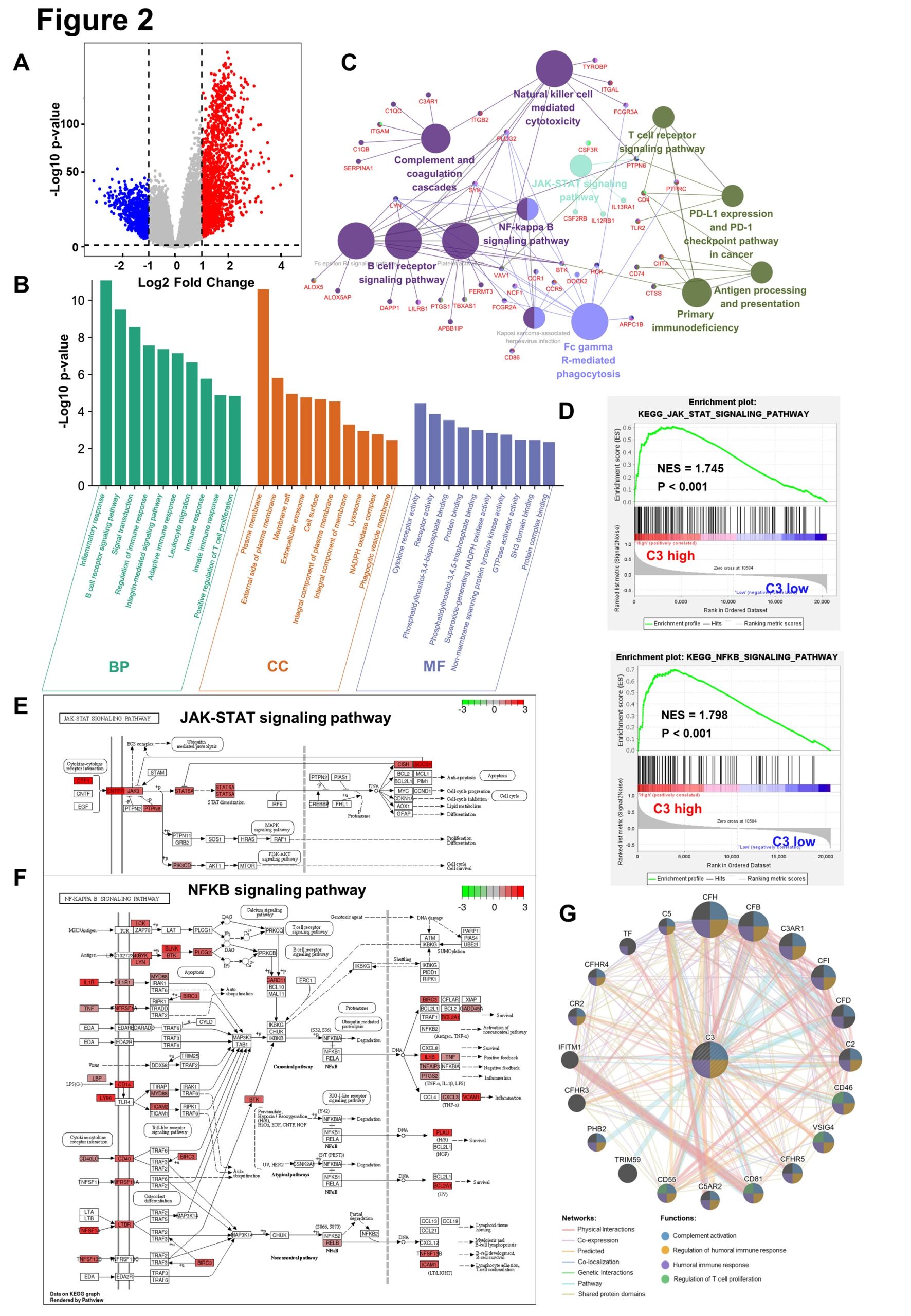
In the TCGA-GBMLGG database, C3 high group and low group do difference analysis, to obtain 1501-regulated genes and 731 down-regulated genes, based on the cut-off criteria (P<0.05 and fold change≥2) (Figure 2A) . The up-regulated genes in the top 200 p-values are selected for GO biological function enrichment analysis. In terms of biological process, C3 up-regulated genes are significantly enriched in the inflammatory reaction and immune response.For cellular components, C3 up-regulated genes are significantly enriched in cell membranes and extracellular matrices. Regarding the molecular function, the up-regulation genes are significantly enriched in cytokine receptor activation (Figure 2B). These significant enrichments can help us further understand the role of C3 in glioma occurrence and progress. Furthermore, KEGG pathway enrichment analysis shows that the C3 up-regulation genes are associated with NF-kB and JAK-STAT signaling pathways (Figure 2C). Similarly, GSEA also reflects that the C3 high expression group up-regulates NF-kB and JAK-STAT signaling pathways (Figure 2D). The KEGG analysis reveals that C3 up-regulated genes increase the expression of the relevant genes of these two signaling pathways, thereby promoting the malignant progression of gliomas (Figure 2E-F). Gene co-expression network is constructed to detect genes showing similar trends (Figure 2G).

Figure 2:  Complement C3 causes malignant progression of glioma through NF-kB and JAK-STAT signaling pathway.

(A). Volcano plot for showing C3 differential expression in TCGA GBMLGG database.(fold change >=2 and p < 0.05).Non-changed genes are shown in gray color. Red color is indicative of up-regulated genes and blue is indicative of down-regulated genes. 

(B). Histogram for showing GO biological function enrichment analyses of C3 up-regulated genes. Biological process enrichment analysis (green), Cell component enrichment analysis(orange) and molecular function enrichment analysis(blue).

(C). Network diagram for displaying KEGG pathway enrichment analyses of C3 up-regulated genes. Large circles represent different signaling pathways, and small dots represent genes enriched in pathways. The lines represent the regulation relationship between genes and pathways.

(D). Gene set enrichment analysis (GSEA) between group high C3 expression and C3 low expression shows NFKB and JAK-STAT signaling pathways.

(E)~(F). Gene expression in the JAK-STAT signaling pathway and NFkB signaling pathway were plotted by PATHVIEW. Red represents up-regulated genes, while green represents down-regulated genes in C3 high expression group.

(G). Construction of gene co-expression networks. The lines represent co-expression networks and the dots represent functions.

Complement C3 Specifically Promotes the Growth of Mesenchymal Subtype Gliomas and Trigger Chemoradiotherapy Resistance

Immunohistochemical staining reveals that C3 is highly expressed in high-grade glioma, especially in area of necrosis (Figure 3A). In the Rembrandt database, C3 mRNA is highly expressed in MES subtype glioma (Figure 3B). C3 high expression and MES subtype correlation signaling pathways are significantly enriched (P<0.01) (Figure 3C). In fact, MES subtype gliomas are highly radiotherapy and chemotherapy resistant. Therefore, we analyze the effect of different treatments on the prognosis of glioma. We found that chemoradiotherapy can significantly prolong the survival of glioma patients (Figure 3D). However, in the group of chemoradiotherapy, C3 high expression might cause chemoradiotherapy resistance in glioma patients and affect their prognosis (Figure 3E).

Figure 3: Complement C3 specifically promotes the growth of mesenchymal subtype gliomas and trigger chemoradiotherapy resistance.

(A). The immunohistochemistry of complement C3 in normal tissues(n=3), low-grade gliomas(n=4), and high-grade gliomas(n=11). The data was downloaded from The Human Protein Atlas. The box plot showing the quantification of the C3 positive rate in different tissues. ‘ns’ represents no significance, *P<0.05.

(B). The box plot shows the C3 mRNA expression level of different glioma subtypes in Rembrandt database. (Non-tumor, n=28; PN, n=76; CL, n=72; MES, n=71). *P<0.05, **P<0.01, ***P<0.001, ****P<0.0001.

(C). Gene set enrichment analysis (GSEA) between group high and low of C3 expression showed signaling pathways of different glioma subtypes in TCGA GBMLGG database.

(D). Kaplan–Meier survival curves for the different treatment groups of the TCGA GBM database (Only Chemotherapy, n=10; Only Radiotherapy, n=53; Chemoradiotherapy, n=324). *P<0.05, **P<0.01, ***P<0.001.

(E). Kaplan–Meier survival curves for the high(n=162) and low(n=162) mRNA expression level of C3 in chemoradiotherapy group(n=324).


Table 1: Multivariable Cox Regression Analysis in C3 expression levels and clinical features. *P<0.05, **P<0.01, ***P<0.001.

Discussion

Gliomas account for approximately 80% of primary central nervous system (CNS) malignant tumors, with high invasiveness, recurrence, high mortality and other characteristics [2]. Currently, the standard treatment for glioma is surgery combined with radiotherapy and chemotherapy [22,23]. In fact, due to the deep location of glioma and chemoradiotherapy resistance, the prognosis of patients remains poor [24,25]. Therefore, in response to these difficulties, we need to propose new treatment options.

Studies have shown that patients with MES signatures belong to the poor prognosis subtype and are resistant to standard treatments [26,27]. In this study, we analyzed that C3 high expression causes chemoradiotherapy resistance in MES subtype gliomas. We also found that C3 high expression promotes chemoradiotherapy resistance in gliomas through NF-kB and JAK-STAT signaling pathways. Similarly, a previous study found that a subset of the PN GSCs undergoes differentiation to a MES state [28] in a TNF-a/ NF-kB-dependent manner with an associated enrichment of CD44 subpopulations and radioresistant phenotypes [29,30]. They further show that the MES signature, CD44 expression, and NF-kB activation correlate with lower radiation response and shorter survival in patients [30].

In summary, the expression of C3 is an important factor affecting the chemoradiotherapy resistance of gliomas. Our data shows that C3 high expression may activate NF-kB and JAK-STAT signaling pathway to promote the chemoradiotherapy resistance of glioma, leading to poor prognosis. Therefore, it is a new therapeutic scheme for targeting C3 and associated signaling pathways to inhibit the chemoradiotherapy resistance of gliomas and improve the prognosis of patients.

More information regarding this Article visit: OAJBGSR

Tuesday 30 November 2021

Influence of Clinical Pathway Implementation on the Outcome of Drug Rehabilitation Program Services

Influence of Clinical Pathway Implementation on the Outcome of Drug Rehabilitation Program Services by Jaya Mualimin in Open Access Journal of Biogeneric Science and Research


Abstract

Patient safety, quality, and efficiency are global issues, therefore hospitals must be able to apply clinical pathways through clinical pathways as the main facilities and infrastructure, especially in services for increasingly acute drug addicts. This study aims to analyze the implementation of clinical pathways for drug rehabilitation program outcomes on 1) clinical quality, 2) cost, 3) readmission, 4) satisfaction, and 5) LOS, at RSJD Atma Husada Mahakam. This type of research uses cross-sectional with observational analytic, data collection through distributing questionnaires to 111 respondents, observation and literature study. The results showed that the clinical quality before and after the implementation of the clinical pathway had a significant effect, but the cost of treatment did not show any significance. There is a positive relationship between readmission and the implementation of clinical pathways, as well as addict satisfaction in the LOS rehabilitation room has a significant effect on treatment time and clinical pathways. A recommendation that the 5 (five) variables mentioned above, apart from being cost-effective, can improve the quality of drug rehabilitation services at RSJD Atma Husada Mahakam Samarinda, so it needs to be maintained

Keywords: Outcome; Quality Clinic; Readmission; Cost, Satisfaction; Length of Stay

Introduction

Drug dependence is a type of chronic recurrent brain disease and must receive therapy and rehabilitation. According to a 2016 BNN survey, the prevalence of drug abuse in East Kalimantan is 3.1% of the population aged 15-49 years. 10% of abusers experiencing health problems need to get medical help. Relapse will affect the quality, quality and patient safety (patient safety). if no preventive measures are taken with organizational policies. The cause of the high number of repeat patients/readmissions is uncontrolled relapse. One indicator of the quality and performance of the RSJD is to reduce the readmission rate, in 2015 the RSJD reduced the readmission rate to only 0.37%. [1].

The hospital implements quality management strategies such as total quality management, quality assurance, and continuous quality management. This strategy tends to focus on management aspects. The following professional aspects will review the definition. Clinical pathway (CP) is an integrated service planning concept that summarizes every step given to patients according to medical service standards, nursing care and other evidence-based services with measurable results. Several researchers have proven the implementation of clinical pathways. Implementation of clinical pathways in opiate therapy patients with methadone, can reduce the cost of therapy and the complications that arise are well controlled. concluded that the implementation of clinical pathways in the therapy of alcoholics, although not related to the length of stay (LOS) but was strongly related to outcome.

Devapriam et al., [2]: implementation of clinical pathways can increase the capacity of per-unit services, shorten the length of stay (LOS), increase the frequency of unit visits, timely assessment of care, and reduce variability in quality. concluded that the implementation of clinical pathways can calculate all types of guarantees or financing for each episode of treatment. Barbieri found significant results in the implementation of clinical pathways for care with a structured organizational approach (clinical governance) from the aspect of quality and low-cost service processes. Cheah said that the implementation of clinical pathways significantly reduces the treatment period and without any side effects or good outcomes. Susi Research, 2009; Chan and Wong, 1999; proves that clinical pathways are associated with increasing cost-effectiveness and significantly reducing the length of treatment.

Several other researchers have found evidence that treatment in one form of regulation (similar to a clinical pathway) can prevent relapse. Prince [3] and Marchisio,proved that making a scheduled treatment similar to a clinical pathway can be used as an indicator of reducing the three-month relapse rate. Before the intervention, the mean of relapse in the intervention group was 30.38% and the control group was 27.54%. After the intervention, there was a 20% reduction in relapse. In the schizophrenic group, 24% had an intervention, while 64% had no intervention [4-10].

Enforcement of clinical pathways for drug dependence Clinical pathways on drug dependence have not been widely studied so that research needs to be done. The implementation of clinical pathways that have been applied, can affect the outcome [11-15]. This study aims to analyze the effect of clinical pathway implementation on service outcomes in the drug rehabilitation program at RSJD Atma Husada Mahakam. The results of the study can be used as an evaluation of the clinical pathway implementation process for the inpatient drug rehabilitation program at RSJD Atma Husada Mahakam. The focus of the research relates to what is in the following picture (Figure 1).

Figure 1: Model Hypothesis

Method

This research method uses a survey model with a quantitative approach that focuses on 111 respondents (patients) who follow an inpatient drug rehabilitation program at Atma Husada Hospital (RSJAH) Mahakam, East Kalimantan, Indonesia [16-18]. The type of data is cross-sectional with analytical observation and literature review. Primary data was obtained from data from clinical pathway forms taken from addicts' medical records and reports on costs for services in drug rehabilitation programs [19-25]. The data analysis technique used the reliability and validity test of the instrument through the SPSS version 22 program, with the data analysis technique in the form of multiple linear regression and associative-causal. This is intended to be able to determine the relationship of influence between the independent variable and the dependent variable through partial and simultaneous tests and prove whether the alternative hypothesis (Ha) is accepted or rejected [25-30].

Research Result

Descriptive Analysis

Based on the results of the study, it was obtained that the description of the status of new patients was still greater than that of old or repeat patients. In (Table 1) in 2015 new patients were 84 addicts (75.7%) and in 2016 new patients were 102 addicts (89.5%). According to the 2016 BNN survey, the prevalence of drug abuse in East Kalimantan is 3.1% of the population aged 15 - 49 years. 10% of abusers experiencing health problems need to get medical help BNN, The prevalence rate of ever used decreased from 8.1% in 2006 to 3.8% in 2016. With the 2012 Accreditation plenary category, the Atma Husada Hospital has an impact on the stigma of mental illness and narcotics [31-35].

Table 1: Distribution of Respondents Characteristics.

Gender of respondents 84.8% are male. The results of this study according to the theory that there are more addicts in men than women, men are more at risk of using drugs than women. The ratio of men to women is about 4 to 1, meaning that among 4 male users there is 1 woman who has tried [36]. The prevalence rate was 13.7% for men and 3.3% for women (2006), while in 2016 it was 6.4% for men and 1.6% for women. The prevalence rate of ever tried using men tends to decrease from 13.7% (2006) to 6.4% (2016). However, in the female group, the trend of decreasing prevalence rates forever and a year using drugs began to be seen from 2009 to 2016 (BNN Survey, 2016).

According to data from the US Substance Abuse and Mental Health Services Administration in 2013, men are more dominant than women in substance abuse of all types and men often overdose and die than women. Including abuse of doctor's prescription. For most age groups, men have a higher level of dependence. However, women can also experience dependence. If women are dependent, the risk is stronger. Robbins et al 1999; Hitschfeld et al 2015; Fox et al 2014; Kennedy et al 2013 and more severe relapses, including women experiencing many legal and social complications including asocial behaviour and abuse. sexual. This problem causes the prevalence of women to be smaller than men [37-45].

Meanwhile, based on age group, table 4.3 was found in 2016 the most age groups were 17-25 late teens group of 115 patients (51.1%) and 26-35 years of early adulthood, 68 patients (30.2%). In 2015, the age group of 17-25 late teens was 58 patients (52.3%). In 2016 at the age of 17-25 late teens as many as 57 patients (50.0%), looking at this number it can be concluded that drug dependence users are dominated by the age group of late teens and early adults. Meanwhile, the education level of most drug rehabilitation patients is junior high school and senior high school, namely 175 (77.7%). In 2015, 2016 consecutively were 80 patients (72.0%), 95 patients (83.3%). However, the large proportion of drug abusers with a bachelor's degree in education does not mean they graduated, some of them are no longer in the campus environment, but are already working in various sectors or even some of them may be unemployed [46-50].

Meanwhile, judging from marital and unmarried status, the most addicted respondents were unmarried status with 121 (53.8%); 56.1% in 2016 and 51.3% in 2015. The place where addicts live is still the most in Samarinda City in 2015 there were 87 patients (78.4%) and in 2016 there were 48 patients (42.1%). In 2016 after the mandatory reporting program (IPWL) and the rehabilitation program for 100,000 addicts, the city of Balikpapan had 19 addicts, as can be seen in Table 1 below

Inferential Descriptive Analysis

LOS Drug Rehabilitation Patient

The average value for the length of stay for drug patients in 2015 or before the implementation of the clinical pathway, which was 42.29 days and in 2016 or after the implementation of the clinical pathway, which was 43.41 days, wherein 2015 it was 74 (66.7%). Meanwhile, in 2016 there were 65 (57.0) less or equal to the average value. The results of the independent sample t-test for LOS obtained p-value = 0.022 < 0.05, which means that there is a significant effect between LOS in 2015 (before clinical pathway implementation) and 2016 (before clinical pathway implementation).

Rehabilitation Patient Fee

The average cost in 2015 was Rp. 14,550,357.87 and in 2016 of Rp. 14,525,219.48, where the number of paying patients is below the mean value of 138 (61.3%). Regulation of the Minister of Health of the Republic of Indonesia. number 50 of 2016 that the cost of drug rehabilitation for a 1-month package is Rp. 6,500,000,000.00. When compared, the Ministry of Health tariff is much lower, if converted to days the Ministry of Health tariff is Rp. 216,666,-/day. the results of the independent sample t-test for the cost of care obtained a value of p = 0.98 > 0.05, which means that there is no significant effect between the cost of care in 2015 (before the implementation of the clinical pathway) and 2016 (before the implementation of the clinical pathway).

Service Quality

There are still a lot of new patient data from old or repeat patients. In 2015 new patients were 84 addicts (75.7%) and in 2016 new patients were 102 addicts (89.5%). From the repeat visit data in 2015 data, addicts who were readmission 2 times were 27 addicts (34.3%) while in 2016; addicts who were treated >2 times there were 12 addicts (10.9%). The results of the independent sample t-test for readmission obtained p-value = 0.006 < 0.05, which means that there is a significant effect between readmission in 2015 (before the implementation of clinical pathways) and 2016 (before the implementation of clinical pathways). Based on the results of the independent sample t-test of the clinical quality variable, the p-value = 0.042, the exit method p = 009 and the visit status p = 006 of the three variables, the p-value = < 0.05, which means a significant influence between clinical quality in 2015 (before implementation of clinical pathways) and 2016 (before the implementation of clinical pathways) [51-57].

Consumer Satisfaction

The satisfaction index of addicts who are being treated in the rehabilitation room is an average of 78.0%. All respondents' satisfaction variables gave satisfying answers above 93.3% to the implementation of clinical pathways asked respondents. The inferential statistics of the findings can be tabulated as follows (Table 2):

Table 2: Bivariate Analysis.

Discussion

With law no. 35 of 2009 on Narcotics article 54 that addicts must be rehabilitated medical and social rehabilitation, with PP no. 25 of 2011 concerning mandatory reporting for drug addicts, following the mandate of the narcotics law that mandatory reporting is the rehabilitation process for addicts at a mandatory reporting institution appointed by the ministry of health and social affairs. Although the diagnosis of drug dependence is included in ICD IX, it is not included in the BPJS health financing, so the government through this mandatory reporting program uses separate financing through the IPWL program. The regulation of the Minister of Health has been revised 3 times (Permenkes No. 50 of 2015) regarding the mandatory reporting technical guidelines for addicts and the procedures for financing and billing inpatient and outpatient rehabilitation costs for addicts.

The purpose of the clinical pathway is an efficient and effective rehabilitation process, reducing variations in procedures and reducing costs. Although cost savings are important, the use of clinical pathways must be evaluated, because the main focus is quality and patient safety. Van Herck et al., 82.5% of the studies reported a positive impact on cost reduction, while 13.5% did not explain the effect and 4% a negative effect.. A 2013 study on the introduction of clinical pathways in postoperative clinical care after major head and neck surgery found a 27% reduction in per-patient costs and several other studies have identified a reduction in length of stay after. Several researchers have proven the implementation of clinical pathways. Ronny Rivani (2014); Implementation of clinical pathways in opiate therapy patients with methadone, can reduce the cost of therapy and the complications that arise are well controlled.

Lacko et. al (2008): concluded that the implementation of clinical pathways can calculate all types of guarantees or financing for each episode of treatment. Barbieri (2009): found significant results in the implementation of clinical pathways for care with a structured organizational approach (clinical governance) from the aspect of quality and low-cost service processes. Cheah (2005): said that the implementation of clinical pathways significantly reduces the treatment period and without any side effects or good outcomes.

The use of clinical pathways has been associated with reduced hospital complications (Rotter et al., 2010) and improved service quality. Van Herck et al. found that 65.5% of the studies reported a positive effect on the outcome, while 32% reported no association with outcome. (Van Herck et al., 2000). Dowdeswell and Yasbeck cited previous studies providing quality and outcome for geriatric patients with depression, (Hindle, Dowdeswell and Yasbeck, 2004). According to Nielsen and Nielsen (2015) that the implementation of clinical pathways in the therapy of alcoholics, although is not related to the length of stay (LOS) but is closely related to the outcome.

The Avalos standard is based on the Minister of Health Regulation No. 50 of 2015 that the 1 monthly package for inpatient rehabilitation treatment is a minimum of 30 days. The average value for the length of stay for drug patients at the Atma Husada Mahakam Mental Hospital in 2015 or before the implementation of the clinical pathway was 42.29 days and in 2016 or after the implementation of the clinical pathway was 43.41 days, wherein 2015 there were 74 (66.7%) while in 2015 2016 as many as 65 (57.0) less or equal to the average value.

Van Herck et al. stated that 62.2% of previous studies had a positive effect on satisfaction, only 29.7% had no effect on satisfaction. Including the research of Renholm, Bryson and Browning who agree that there is an improvement in patient satisfaction. One of the potential benefits of the care pathway is to improve communication between professionals. While one study revealed that although integrated clinical pathways resulted in better health care trust, there was little evidence to suggest that interpersonal relationships and communication needed to be improved, although there was no measurable improvement. On the other hand, based on clinical results, interprofessional communication improved. Interdisciplinary teamwork can be supported by clinical pathways in other fields (Figure 2).

Figure 2: Research Model Results.

Conclusion

In this study, three important things can be found, first, the clinical quality which consists of a) patients running away, b) forced discharge, and c) readmission is highly dependent on the implementation of clinical pathways, secondly, the implementation of clinical pathways has no connection at all with costs. drug rehabilitation treatment, and thirdly the application of clinical pathways makes an important contribution to the satisfaction of patients being treated for drug rehabilitation. This means that the satisfaction factor makes an important contribution to drug rehabilitation services at the Atma Husada Mahakam Hospital.

This means that the service outcomes provided by the Atma Husada Mahakam Hospital are generally good. This can be shown by the number of high satisfaction responses from respondents to each research variable. Similarly, the service quality variable from the five dimensions has a positive and significant influence on customer satisfaction. This reinforces the previous theory that the provision of quality services can certainly create satisfaction for everyone, including the treatment of drug rehabilitation patients.

More information regarding this Article visit: OAJBGSR