Tuesday 26 April 2022

Wilms’s Tumor Gene Mutations: Loss of Tumor Suppresser Function: A Bioinformatics Study by Uzma Jabbar in Open Access Journal of Biogeneric Science and Research

Wilms’s Tumor Gene Mutations: Loss of Tumor Suppresser Function: A Bioinformatics Study by Uzma Jabbar in Open Access Journal of Biogeneric Science and Research 


ABSTRACT

Introduction: Mutation in the Wilms’s Tumor (WT1) gene product has been detected in both sporadic and familial cases suggesting that alteration in WT1 may disrupt its normal function. The study aims to find the protagonist amino acid in WT1 proteins by mutating these residues with other amino acids.

Material and Methods: The 3D modeling approach by MODELLER 4 was utilized to build a homology of WT1 proteins. Quality of the WT1 model was verified by predicting 10 models of WT1 and hence selecting the best one. Stereochemistry of model was evaluated by PROCHECK. Mutational studies were done by WHAT IF. Five human WT1 mutations were modeled which were Lys371→Ala371, Ser415→Ala415, Cys416→Ala416, His434→Asp434 and His434→Arg434.

Result: Based on active side of WT1 protein and its role in DNA binding mutation. No significant change was observed when Lys371 was mutated to Ala371, Ser415 was mutated to Ala415. Significant change was observed in Cys416 mutated to Ala416. In mutant Ala416, loss of coordination with the metal ion Zn was also predicted. In case of Mutants His434→Asp434, there was a loss of coordination of metal ion (Zn203) with mutant Asp434. In case of mutant His434→Arg434, there was a loss of Zn203 coordination with Arg434. His434 does not interact directly with any DNA base, whereas mutated Arg434 is predicted to interact directly with DNA base.

Conclusion: It is concluded that mutation of amino acid residue Cys416→Ala416, His434→Asp434 and His434→Arg434 may lose the proto-oncogenic function of WT1.


Keywords: WT1 protein, MODELLER9.0, Mutation, Active side residues

Introduction

WT1, is a protein, which in humans is encoded by the WT1 gene on chromosome 11p13. The WT1 is responsible for the normal kidney development.  Mutations in this gene are reported to develop tumors and developmental abnormalities in the genitourinary system. Conversion of proto-oncogenic function to oncogenic in WT1 has also been documented cause of various hematological malignancies. (***)

Multifaceted protein of WT1 gene has transcriptional factor activity [1]. It regulates the expression of insulin-like growth factor and transforming growth factor system, implicated in breast tumorigenesis [2]. A main function of WT1 is to regulate transcription, which control the expression of genes involved in the process of proliferation and differentiation [3]. In wide range of tumor, WT1 is shown to be predisposing factor for cancer, therefore it has become hot target in research to find out it’s inhibitor which can be safely used as a treatment of cancer. It can induce apoptosis in embryonic cancer cell, presumably through the withdrawal of required growth factor survival signal [4]. WT1 is involved in the normal tissue homeostasis and as an oncogene in solid tumors, like breast cancer [5]. Increased expression of WT1 is related with poor prognosis in breast cancer6. A number of hypotheses are postulated for the relationship of WT1 with tumorigenesis. Acceding to one of the hypothesis, elevated levels of WT1 in tumors may be related with increased proliferation because normally WT1 have a role with apoptosis [7,8]. Another study proposed that WT1 can alter many genes of the the family of BCL2 [9,10] and also have a role to regulate with Fas-death signaling pathway [11]. Furthermore, it is suggested that WT1 can encourage cell proliferation by up-regulation of protein cyclin D1 [12].

A group of workers hypothesized that WT1 has been observed in the vasculature of some tumour types [13]and its expression may be related with angiogenesis especially in endometrial cancer [14]. Another hypothesis based on the fact that WT1 is a main regulator of the epithelial/mesenchymal balance and may have a role in the epithelial-to-mesenchymal transition of tumor cells [3]. Expression of WT1 is higher in estrogen receptor (ER) positive than in ER negative tumors. It is therefore possible that WT1 not only interact with ER alpha, but it may orchestrate its expression [15]. A study, on triple negative breast cancers [7] has shown that high WT1 levels associate with poor survival due to increased angiogenesis [16,17], altered proliferation/apoptosis10,11, and induction of cancer- epithelial-to-mesenchymal transition4. In breast tumors, WT1 is mainly related with a mesenchymal phenotype and increased levels of CYP3A4 [18]. A mutation in the zinc finger region of WT1 protein has been identified in the patients that abolished its DNA binding activity [19]. A study also observed that the mutation in the WT1 gene product has been detected in both sporadic and familial cases suggesting that alteration in WT1 may disrupt its normal function [20].  Bioinformatics approaches are being utilized to resolve the biological problems. Efforts start with the prediction of 3D structures. To achieve the aim, study was designed to view 3D structure of WT1, tumor suppressor protein predicted by homology modeling and to study the role of crucial residues in WT1 proteins by mutating these residues with other amino acids.

Material and Methods

3D structure of WT1 was taken as target of human WT1. Figure 1 shows the normal interaction of WT1 with DNA strands based on the crystal structure of a zinc finger protein.

Figure 1: Homology model of the C-terminal fragment of Human Wilms Tumor protein with bound DNA strands based on the crystal structure of a zinc finger protein, znf268, (PDB; id:1aay) and a five-finger protein, GLI (PDB; id:2gli).

The binding of protein-DNA complex involves four zinc finger binding domain in the C-terminal/Zn finger region of WT1. These are;

  • Cys325, Cys330 and His339, His343: figure 2; yellow highlighted.
  • Cys355, Cys360 and His373, His377: figure 3; yellow highlighted.
  • Cys385, Cys388 and His397, His401: figure 4; yellow highlighted.
  • Cys416, Cys421 and His434, His435: figure 5; yellow highlighted.

Figure 2

Figure 3

Figure 4

Figure 5

The 449 amino acid sequences of WT1 were used for homology modeling. Sequences of WT1 were retrieved from Swiss Prot Data Bank in FASTA format [21]. The best suitable templates were used for 3D-structure prediction. The retrieved amino acid sequences of WT1 were subjected to BLAST [22]. Templates were retrieved on the base of query coverage and identity. The 3D structures were predicted by MODELLER 9.0 [23] that is the requirement of 3D structure building of target protein. Tools including stereochemistry and Ramchandran plots were used for the structure evaluation [24]. Identification of Template was carried out, and Sequence Alignment was carried out by using FASTA, BLAST. Quality of the WT1 model was verified. Stereochemistry of model was evaluated by PROCHECK [25]. Mutational studies were done by WHAT IF [26]. Five human WT1 mutants are modeled. These were: Lys371→Ala371, Ser415→Ala415, Cys416→Ala416, His434→Asp434 and His434→Arg434.

Results and Discussion

The study was largely based on active side of the WT1 and its role in DNA binding mutation. Zinc finger binding domain interact selectively and non-covalently. This zinc finger-binding domain is the classical zinc finger domain, in which two conserved cysteine and histidine co-ordinate a zinc ion at the active site.

Cys416®Ala416 MUTANT

Significant change was observed in Cys416 mutated to Ala416. In mutant Ala416, reduction in the Van der Waal’s contact between the amino acids. Loss of coordination with the metal ion Zn was also predicted (Figure 6 A and B).

                                                       Figure 6A                                                                   Figure 6B

Figure 6 A and B: Wild Type (Cys416) and mutated (Ala416) WT1.  Distance between Zn and Cys416 is increased in mutated (Ala416) model. Cys416 is predicted to be found in the vicinity of His434 and His438 which are implicated in catalysis (6A) while Ala416 can only interact with His434 and not with His438 in the mutated model (6B).

Cys416 is located at the domain interface with its polar side chain completely buried (0.00 Å). Replacement of this amino acid may account for considerable changes in the interior of protein (Table 1). We have predicted the possible changes that arise due to the mutation of Cys to Ala by molecular modeling experiments. Amino acids, Pro419, Ser420, Cys421, His434 and some atoms of His438 (ND1, NE2, CD2 and CE1) are present near Cys416. Zinc (Zn203) is also present in the vicinity (1.82 Å) of Cys416 (Figure 6). The mutated residue, Ala is also predicted to remain buried (0.00 Å) in the interior of protein. Significant change is observed however, in the surrounding area of the mutated Ala416. Only a few atoms of His434 (CD2 and NE2) and His438 (CE1) were seen in the surrounding. This may reduce the Van der Waal's contacts between the respective amino acids. The loss of coordination with the metal ion, zinc was also predicted as the distance is increased from 1.82 Å to3.12 Å.   It is therefore predicted that Cys416 plays a vital role in the interaction with other amino acid residues as well as in the metal coordination. It is observed that there is a possibility of loss of these interactions in case of Cys416 replacement.

His434®Arg434 MUTANTS

In case of mutant His434→Arg434, there was a loss of zn203 coordination with Arg434.  His434 does not interact directly with any DNA base, whereas mutated Arg434 is predicted to interact directly with DNA base, A1. This suggests that change might effect on the DNA binding pattern, Figure 7 A and B.

                                                       Figure 7A                                                                   Figure 7B

FIGURE 7 A and B: Wild Type (His434) and mutated (Arg434) WT1.  Distance between Zn and His434 is increased in mutated model. Arg434 is predicted to bind DNA base A1 (B) while His434 in the original model (A) show no bonding with DNA base.

In case of mutation of His434®Arg434, the distance between the mutated Arg and zinc (Zn203) was increased from 2.28 Å to5.00 Å suggesting that there could be a loss of coordination with the metal ion. Mutational studies proved that hydrogen bonding network close to the zinc-binding motif plays a significant role in stabilizing the coordination of the zinc metal ion to the protein23. The mutated amino acid, Arg434 also moved considerably form buried to relatively exposed environment (2.28 Å to 5.35 Å). Presence of positively charged Arg on the surface could account for additional interaction of the protein with other proteins or with the surrounding water molecules. His434 does not interact directly with any DNA base whereas mutated Arg434 is predicted to interact directly with DNA base Adenine, A1. (Figure 7). This suggests that the change might cause the DNA binding pattern.

TABLE 1: Comparison of surface accessibilities (Å) of the wild type and mutated residues and those in the vicinity of the mutated residues in the five WT1 mutants

Lys371®Ala371 and Ser415®Ala415 MUTANT

No significant change was observed when Lys371 was mutated to Ala371, and Ser415 was mutated to Ala415. It is observed in this mutation that the change that arise in the overall structure and surrounding amino acid residues (Table 1). Lys371 is present on the surface (accessibility = 47.04 Å) of the WT1 molecule. It was observed that the internal protein structure was not affected considerably, as Lys371 is present on the outer most surface of the protein. In the original model, Lys371 stacks against thymine. It also forms a water-mediated contact with side chain hydroxyl of Ser367. Although, Ala371 also stacks against the same DNA base but the distance is slightly altered. The hydrogen bond between Ala371 and Ser367 has not been predicted in the mutated model.  It has been demonstrated that mutation within finger 2 and 4 abolished sequence specific binding of WT1 to DNA bases19. The mutation of the corresponding lysine in a peptide could reduce its affinity for DNA seven folds [27].  On the other hand, it is reported [28] that a surface mutation would not cause a significant change in the internal structure of protein.  However, the replacement of a basic polar residue with a non-polar one could account for a reduction in polarity.  The modeling studies of Lys to Ala mutation do not however support this finding and require further analysis.

Mutation of Ser415®Ala415 in the WT1 model (Table 1). Ser415 is located near the active center of WT1. It has been demonstrated that Ser415 makes a water-mediated contact with phosphate of DNA base, guanine [20]. In our predicted model of WT1, Ser415 makes two water (numbers 516 and 568) mediated contacts. Mutation of this Ser with Ala resulted in the loss of one of these contacts leading to the loss of binding. The replacement of relatively polar residue, Ser to a non-polar one, Ala could account for this reduced interaction. This is also evident by a slight decrease in the accessibility of Ala (Ser415 = 7.96 Å; Ala415 = 7.61 Å).

His434®Asp434 MUTANTS

In case of mutants His434→Asp434, there was a loss of coordination of metal ion (zn203) with mutant Asp434. Glu430 move from relatively exposed to completely buried environment. His434 is also present at the active center of WT1. We predicted two mutants; His434®Asp434 and His434®Arg434 mutants by molecular modeling (Table 1). In case of His434®Asp434 mutation, the water mediated contact is lost. The distance between mutated Asp and zinc (Zn203) was also increased from 2.28 Å to 3.57 Å suggesting that there could be a loss of coordination with the metal ion as well. The amino acid Glu340 that is present near His434 also moved considerably form relatively exposed to completely buried environment (14.83 Å to 00.0 Å).

Conclusion

It is concluded that mutation of amino acid residue Cys416→Ala416, His434→Asp434 and His434→Arg434 of WT1 may lose its function to regulate the function of genes by binding to specific parts of DNA. Besides the mutation of above-mentioned amino acid residue, the role of WT1 in cell growth, cell differentiation, apoptosis and tumor suppressor function is also lost.

More information regarding this Article visit: OAJBGSR


https://biogenericpublishers.com/pdf/JBGSR.MS.ID.00250.pdf
https://biogenericpublishers.com/jbgsr-ms-id-00250-text/

Tuesday 12 April 2022

Gene Editing via Integrase Enzyme by Umair Masood* in Open Access Journal of Biogeneric Science and Research

Gene Editing via Integrase Enzyme by Umair Masood* in Open Access Journal of Biogeneric Science and Research 


Short Communication

Targeted integrase enzyme is a most powerful tool for mediating genome alteration with high precision. The gene of the interest is directly catalyze nucleophilic attack of the 3 prime hydroxyl group at the end of processed DNA on a pair of phosphodiaster bond in the targeted DNA or genome of the interest. Integrase gene editing method contain two parts gene of the interest with integrase enzyme and desire genome. The integrase enzyme is start integration the gene of the interest into the desire genome or DNA.

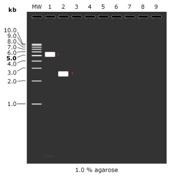
Results via Gel electrophoresis

In order to check that whether gene of interest is integrated or not we can perform a gel electrophoresis. The gel contain a two band and B.A band is a vector of human insulin vector which can show the negative control and band is a vector of human insulin but the gene have some sequence mutation [1-5]. We can add a gene of the stranded human insulin by using integrase enzyme and gene can be precisely add to the B band that why B band is in 3.0 and A band is 6.0 which mean that A band is high molecular weight than the B band and a Marker must be 1kb and agarose should be 1% [6-9].


More information regarding this Article visit: OAJBGSR


https://biogenericpublishers.com/pdf/JBGSR.MS.ID.00249.pdf
https://biogenericpublishers.com/jbgsr-ms-id-00249-text/

Wednesday 6 April 2022

Giant Cell Tumor of the Distal Tibia and Fibula (Rare Location) by Mohamed Hamid Awadelseid* in Open Access Journal of Biogeneric Science and Research


Giant Cell Tumor of the Distal Tibia and Fibula (Rare Location) by Mohamed Hamid Awadelseid* in Open Access Journal of Biogeneric Science and Research 

ABSTRACT

Giant cell tumor of the distal tibia is a rare, benign and usually asymptomatic condition. The discovery is sometimes made following a medical imaging examination or a painful symptom or more often a visible or palpable swelling with or without vascular and/or nerve compression. At an advanced stage, the X-ray is of paramount importance. The well complete surgical resection is part of the therapeutic. We present a clinical case report of a young man with a giant cell tumor localized in the distal tibia in Khartoum, Sudan. This case concerns a 37-year-old patient who presented in July 2021 of a huge painful swelling at left distal tibia treated with bonesetter at Kassla, eastern Sudan and whose X-ray radiography showed lytic lesion of the cortical bone in the lower third of the tibia. After the operative resection of the tumor mass, the pathological examination of the operative specimen revealed the diagnosis of a giant cell tumor. A giant cell tumor is a benign condition, with a few symptoms and the location at the ankle is exceptional. Complete surgical resection is a viable treatment option.

KeywordsGiant Cell Tumor, Wide Surgical Resection, tibia

Introduction

Giant cell tumor (GCT) of bone is one of the commonest benign bone tumors encountered by an orthopedic surgeon. The reported incidence of GCT in the Oriental and Asian population is higher than that in the Caucasian population and may account for 20% of all skeletal neoplasms. It has a well-known propensity for local recurrence after surgical treatment Although considered to be benign tumors of bone [1].

GCT has a relatively high recurrence rate. Metastases occur in 1% to 9% of patients with GCT and some earlier studies have correlated the incidence of metastases with aggressive growth and local recurrence. Current recurrence rates between 10-20% with meticulous curettage and extension of tumor removal using mechanized burrs and adjuvant therapy are a vast improvement on the historically reported recurrence rates of 50-60% with curettage alone. GCT of bone constitutes 20% of biopsy analyzed benign bone tumors. It affects young adults between the ages of20 and 40 years, several authors have reported a slight predominance of women over men. However, GCT can be seen in patients over 50 years old. Ninety percent of GCT exhibits the typical epiphyseal location. Tumor often extends to the articular subchondral bone or even abuts the cartilage. The joint and or its capsule are rarely invaded. In rare instances in which GCT occurs in a skeletally immature patient, the lesion is likely to be found in the metaphysis,The most common locations, in decreasing order are the distal femur, the proximal tibia, the distal radius, and the  sacrum .Fifty percent of GCTs arise around the knee region. Other frequent sites include the fibular head, the proximal femur, and the proximal humerus. Pelvic GCT is rare[6]. Multicentricity or the synchronous occurrence of GCT in different sites is known to occur, but is exceedingly rare [2].

We report a case of a 37-year-old man who presented in July 2021, with a huge painful swelling his left distal tibia with an X-ray radiography showing lysis of the cortical bone in the lower third of the tibia. After the operative excision of the tumor mass, the pathological examination of the specimen revealed the diagnosis of a giant cell tumor. With a lytic lesion of the distal tibia and fibula bones in a young man on X-ray, one must think of a giant cell tumor [3].

Case Report

We report the case of a 37-year-old young man with no notable pathological antecedents who presented at the orthopedic consultation for a painful swelling of the left distal tibia and fibula, that had been evolving for 5 months and without any alteration of the general state. There was change in color and consistency of the skin with respect to the tumor action.

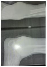
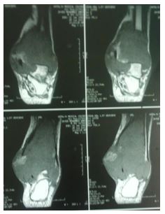
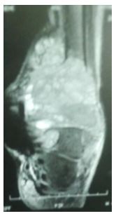
On physical examination, there was a dorsoflextion blockage of foot because of the large volume occupied by the tumor mass and the articular destruction at the level of the distal tibio-fibular joint; plantar flexion estimated at 10˚ and dorsal flextion at 3˚; inversion at 5˚ and eversion at 2˚. The x-ray showed a lesion with blurred boundaries, extending into the soft tissue that is not limited by a bony shell; with destruction of the cortex, invasion of soft parts and honeycomb pseudo-partitions (Figure 1-2). And finally, the Magntic Resonant Imaging (MRI) of the left leg showed (Figure 3-4-5-6) a lysis of the cortex of the lower extremity of the leg. It corresponds to grade 3 of the Campanacci and Merled ’Aubignee classification.

Figure 1: X.ray Show lytic lesion in the cortical bone involve distal tibia and fibula with cortical break posteriomedially with blurred boundaries, extending into the soft tissue that is not limited by a bony shell; with destruction of the cortex, invasion of soft parts.

Figure 2: Show X.ray of lytic lesion in cortical bone involve distal tibia and fibula with cortical break posteri-omedially with blurred boundaries, extending into the soft tissue that is not limited by a bony shell; with destruction of the cortex, invasion of soft parts.

Figure 3: MRI report demonstrates heterogeneous lesion involve distal tibia and fibula.

Figure 4: MRI Show heterogeneous lesion involve distal tibia and fibula.

Figure 5: MRI Show heterogeneous lesion involve distal tibia and fibula with cortical break posteriomedially.

Figure 6: MRI Show heterogeneous lesion involve distal tibia and fibula with fatty infiltration and enhancement of blood vessel at medial site.

A complete surgical resection below knee amputation (BKA) was offered to the patient. Under spinal anesthesia, this incision was made proximally to expose a healthy portion of the leg bone. Surgical removal of the tumor by BKA a proximal resection of the tibia and fibular bone by about 2 cm in the healthy zone. The anatomopathological assessment (Figure7) showed abundant mononuclear cells and discrete nuclear anomalies with marked mitotic activity, but without atypical forms. The histological examination of the bone fragments confirmed a grade 3 giant cell tumor according to Sanerkin, Jaffe Lichtenstein and Pottis. CT scan was done to exclude pulmonary metastases (Figure 8). At three weeks removal of suture and start physiotherapy of knee. Surgical treatment with the excision of the large tumor mass by BKA improved the function of the leg and general condition of the patient.

Figure 7: Histopathology report demonstrates giant cell tumor  involve distal tibia and fibula wit safety margin.

Figure 8: CT scan report demonstrates normal scan of chest exclude pulmonary metastases.

Discussion

Giant cell tumors (GCT) account for 5%–9% of all benign and malignant bone tumors. They are considered benign but may present a progressive, potentially malignant clinical course. GCT recur in a high percentage of cases, become sarcomatous, yet produce metastases even without apparent malignant changes [4].

In the literature, the recurrence rate varies considerably, depending not only on the site and extension of the lesion but also on the type of primary treatment performed [5]. Successful treatment of GCTs and the adequacy of tumor removal is influenced by tumor location ,associated fracture, soft tissue extension ,and understanding of the functional consequences of resection .each option has advantage and disadvantage [6]. This technique has the advantage of preserving knee joint function, improved general and psychological condition of the patient and recurrences are no more frequent than with other techniques according to several authors.  Excision with tumor free margins is associated with lesser recurrence rates. However, for periarticular lesions this is usually accompanied with a suboptimal functional outcome [7]. Various studies suggest that wide resection is associated with a decreased risk of local recurrence when compared with intralesional curettage and may increase the recurrence free survival rate from 84% to 100% (8). However, wide resection is associated with higher rates of surgical complications which led to functional impairment, generally necessitating reconstruction.  This procedure resulted in good function preserving knee joint function after removal of suture 3 weeks later. After 5 months follow up, there was no recurrence or functional sequelae of the leg. Giant cell bone tumors generally have a good prognosis [9].

Conclusion

The giant cell tumor of the distal tibia and fibula bone, although rare, does not present any particularity. The gold standard X-ray image guided radiography with the bone tissue histology confirmed the diagnosis. use a CT scan or an MRI study if we fear an invasion of soft parts. Surgical treatment preserved joint function. This should be considered when presented with a lytic epiphyseal bone lesion.

More information regarding this Article visit: OAJBGSR


https://biogenericpublishers.com/pdf/JBGSR.MS.ID.00248.pdf
https://biogenericpublishers.com/jbgsr-ms-id-00248-text-2/

Friday 1 April 2022

Contribution of Award-Winning Research to the Visibility of Science on a University by Raquel de la Cruz Soriano in Open Access Journal of Biogeneric Science and Research

Contribution of Award-Winning Research to the Visibility of Science on a University by Raquel de la Cruz Soriano in Open Access Journal of Biogeneric Science and Research 


ABSTRACT

This article presents the experience in the preparation and presentation of results to the call for awards of the Ministry of Science, Technology and Environment, evidence of the impact achieved. The methods of science were applied such as: analysis and synthesis, historical and logical, hypothetical deductive; within the empirical ones: observation, document review, interview, training actions, conducting scientific sessions, giving conferences and joint elaboration. The scientific results with the potential to opt for the call were determined by reviewing documents, interviewing teachers-researchers, holding scientific sessions on the subject and giving lectures on the methodological procedure.

KEYWORDS: Science and Technological innovation; Prizes; Scientific results.

Introduction

This article highlights one of the indicators of the Science, Technology and Innovation process as one of the pillars of the management of substantive processes in municipal university centers. As innovation is a social process, the inadequacies of the educational system and high levels of poverty and social inequality affect the development and performance of innovation systems. The links between universities and public Research + Development (R + D) centers, with the productive sector, are mostly based on obtaining information and training and not on forms of interaction to reverse specific problems, through the application of results. scientists, that is, closing the research cycle [1]. The evaluation of the impact of science and technology constitutes a strategic need, as a way to verify the development of a country, its scientific policy, as well as its management in terms of society and the human beings who live in it [9]. Given the new development perspectives depending on the local, [8] indicates the need for greater integration of the Municipal University Centers with the productive sector, increasing the connections between the different actors outside the municipality, which enhances the development of the learning and innovation, from the increase of the scientific debate in the locality, which means increasing the rigor in the analysis of the problems [7]. sustains that the research bases a broad innovation approach or “DUI innovation mode” (doing, using, interacting) in which learning is key [5-6]. He argues in favor of innovation systems that favor social inclusion and care for the environment. Technological trajectories should benefit the human groups involved, expand their knowledge, improve their quality of life, among others [3].

Achieving relevance in science and technology has its starting point in the needs of economic, social and cultural development of the territory, these guide the substantive processes of science, technique and postgraduate that are executed in the Municipal University Centers (CUM); In these, the search is encouraged based on the need to possess knowledge and they drive action. The learning needs determine the high-level continuing training activities, based on the demand of the group of professionals graduated from a territory, who become the main clients of the postgraduate studies of their university. This activity enhances interaction with the university environment and provides new knowledge to those involved in research and development projects, in turn, it is a search for new research needs and opportunities, research projects; so that an ascending cyclical and interactive process is established.

On many occasions, quality in education centers has been related to terms such as: prestigious centers or centers of excellence, certified or accredited, with good economic resources and good infrastructures or good facilities, centers with excellent academic results, with good teachers and great leaders, with the satisfaction of parents and students and with evaluation of all kinds: of the system, of the educational processes, of the results.

As part of the dynamics of science and technological innovation management, the scientific visibility indicators express the relevance and relevance of the research results. That is why achieving territorial recognition is a goal of the Cabaiguán CUM, an aspect that has been projected in the strategic planning of the institution in different stages, which has required the execution of different training actions for the faculty.

This article proposes to expose the experience in the preparation and presentation of results to the call for awards of the Ministry of Science, Technology and Environment, evidence of the impact achieved.

Figure 1: Mapa de proceso, tipo de proceso: Ciencia e Innovación Tecnológica. Fuente: FUM (2014) b)

Figura2: Diagrama de flujo del proceso de Ciencia e Innovación Tecnológica. Fuente: elaboración propia.


Table1: Premios otorgados por el CITMA (2009-2020)

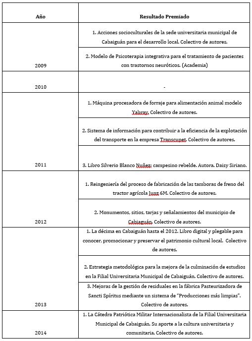
Table2: Trabajos premiados por el CITMA.

Conclusion

The increase in results in the calls made as of 2012 is significant, determined mainly by the results of investigations of teachers in the completion of master's thesis, the completion of study of undergraduate students, innovations in companies in the territory and in university processes; as part of research projects or problem bank in local institutions. The experience presented shows the viability of the flow diagram for the management of science and innovation in a university campus, where the training carried out to the faculty and the method of joint preparation of the files to be presented to the awards calls, allow materialize presented results.

More information regarding this Article visit: OAJBGSR


https://biogenericpublishers.com/pdf/JBGSR.MS.ID.00247.pdf
https://biogenericpublishers.com/jbgsr-ms-id-00247-text-2/