Giant Cell Tumor of the Distal Tibia and Fibula (Rare Location) by Mohamed Hamid Awadelseid* in Open Access Journal of Biogeneric Science and Research
ABSTRACT
Giant cell tumor of the distal tibia is a rare, benign and usually asymptomatic condition. The discovery is sometimes made following a medical imaging examination or a painful symptom or more often a visible or palpable swelling with or without vascular and/or nerve compression. At an advanced stage, the X-ray is of paramount importance. The well complete surgical resection is part of the therapeutic. We present a clinical case report of a young man with a giant cell tumor localized in the distal tibia in Khartoum, Sudan. This case concerns a 37-year-old patient who presented in July 2021 of a huge painful swelling at left distal tibia treated with bonesetter at Kassla, eastern Sudan and whose X-ray radiography showed lytic lesion of the cortical bone in the lower third of the tibia. After the operative resection of the tumor mass, the pathological examination of the operative specimen revealed the diagnosis of a giant cell tumor. A giant cell tumor is a benign condition, with a few symptoms and the location at the ankle is exceptional. Complete surgical resection is a viable treatment option.
Keywords: Giant Cell Tumor, Wide Surgical Resection, tibia
Introduction
Giant cell tumor (GCT) of bone is one of the commonest benign bone tumors encountered by an orthopedic surgeon. The reported incidence of GCT in the Oriental and Asian population is higher than that in the Caucasian population and may account for 20% of all skeletal neoplasms. It has a well-known propensity for local recurrence after surgical treatment Although considered to be benign tumors of bone [1].
GCT has a relatively high recurrence rate. Metastases occur in 1% to 9% of patients with GCT and some earlier studies have correlated the incidence of metastases with aggressive growth and local recurrence. Current recurrence rates between 10-20% with meticulous curettage and extension of tumor removal using mechanized burrs and adjuvant therapy are a vast improvement on the historically reported recurrence rates of 50-60% with curettage alone. GCT of bone constitutes 20% of biopsy analyzed benign bone tumors. It affects young adults between the ages of20 and 40 years, several authors have reported a slight predominance of women over men. However, GCT can be seen in patients over 50 years old. Ninety percent of GCT exhibits the typical epiphyseal location. Tumor often extends to the articular subchondral bone or even abuts the cartilage. The joint and or its capsule are rarely invaded. In rare instances in which GCT occurs in a skeletally immature patient, the lesion is likely to be found in the metaphysis,The most common locations, in decreasing order are the distal femur, the proximal tibia, the distal radius, and the sacrum .Fifty percent of GCTs arise around the knee region. Other frequent sites include the fibular head, the proximal femur, and the proximal humerus. Pelvic GCT is rare[6]. Multicentricity or the synchronous occurrence of GCT in different sites is known to occur, but is exceedingly rare [2].
We report a case of a 37-year-old man who presented in July 2021, with a huge painful swelling his left distal tibia with an X-ray radiography showing lysis of the cortical bone in the lower third of the tibia. After the operative excision of the tumor mass, the pathological examination of the specimen revealed the diagnosis of a giant cell tumor. With a lytic lesion of the distal tibia and fibula bones in a young man on X-ray, one must think of a giant cell tumor [3].
Case Report
We report the case of a 37-year-old young man with no notable pathological antecedents who presented at the orthopedic consultation for a painful swelling of the left distal tibia and fibula, that had been evolving for 5 months and without any alteration of the general state. There was change in color and consistency of the skin with respect to the tumor action.
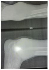
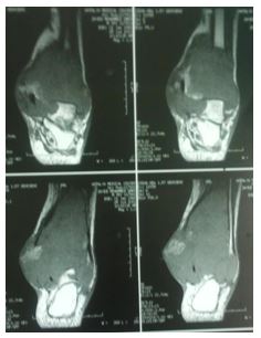
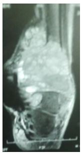
On physical examination, there was a dorsoflextion blockage of foot because of the large volume occupied by the tumor mass and the articular destruction at the level of the distal tibio-fibular joint; plantar flexion estimated at 10˚ and dorsal flextion at 3˚; inversion at 5˚ and eversion at 2˚. The x-ray showed a lesion with blurred boundaries, extending into the soft tissue that is not limited by a bony shell; with destruction of the cortex, invasion of soft parts and honeycomb pseudo-partitions (Figure 1-2). And finally, the Magntic Resonant Imaging (MRI) of the left leg showed (Figure 3-4-5-6) a lysis of the cortex of the lower extremity of the leg. It corresponds to grade 3 of the Campanacci and Merled ’Aubignee classification.

Figure 1: X.ray Show lytic lesion in the cortical bone involve distal tibia and fibula with cortical break posteriomedially with blurred boundaries, extending into the soft tissue that is not limited by a bony shell; with destruction of the cortex, invasion of soft parts.

Figure 2: Show X.ray of lytic lesion in cortical bone involve distal tibia and fibula with cortical break posteri-omedially with blurred boundaries, extending into the soft tissue that is not limited by a bony shell; with destruction of the cortex, invasion of soft parts.

Figure 3: MRI report demonstrates heterogeneous lesion involve distal tibia and fibula.

Figure 4: MRI Show heterogeneous lesion involve distal tibia and fibula.

Figure 5: MRI Show heterogeneous lesion involve distal tibia and fibula with cortical break posteriomedially.

Figure 6: MRI Show heterogeneous lesion involve distal tibia and fibula with fatty infiltration and enhancement of blood vessel at medial site.
A complete surgical resection below knee amputation (BKA) was offered to the patient. Under spinal anesthesia, this incision was made proximally to expose a healthy portion of the leg bone. Surgical removal of the tumor by BKA a proximal resection of the tibia and fibular bone by about 2 cm in the healthy zone. The anatomopathological assessment (Figure7) showed abundant mononuclear cells and discrete nuclear anomalies with marked mitotic activity, but without atypical forms. The histological examination of the bone fragments confirmed a grade 3 giant cell tumor according to Sanerkin, Jaffe Lichtenstein and Pottis. CT scan was done to exclude pulmonary metastases (Figure 8). At three weeks removal of suture and start physiotherapy of knee. Surgical treatment with the excision of the large tumor mass by BKA improved the function of the leg and general condition of the patient.

Figure 7: Histopathology report demonstrates giant cell tumor involve distal tibia and fibula wit safety margin.

Figure 8: CT scan report demonstrates normal scan of chest exclude pulmonary metastases.
Discussion
Giant cell tumors (GCT) account for 5%–9% of all benign and malignant bone tumors. They are considered benign but may present a progressive, potentially malignant clinical course. GCT recur in a high percentage of cases, become sarcomatous, yet produce metastases even without apparent malignant changes [4].
In the literature, the recurrence rate varies considerably, depending not only on the site and extension of the lesion but also on the type of primary treatment performed [5]. Successful treatment of GCTs and the adequacy of tumor removal is influenced by tumor location ,associated fracture, soft tissue extension ,and understanding of the functional consequences of resection .each option has advantage and disadvantage [6]. This technique has the advantage of preserving knee joint function, improved general and psychological condition of the patient and recurrences are no more frequent than with other techniques according to several authors. Excision with tumor free margins is associated with lesser recurrence rates. However, for periarticular lesions this is usually accompanied with a suboptimal functional outcome [7]. Various studies suggest that wide resection is associated with a decreased risk of local recurrence when compared with intralesional curettage and may increase the recurrence free survival rate from 84% to 100% (8). However, wide resection is associated with higher rates of surgical complications which led to functional impairment, generally necessitating reconstruction. This procedure resulted in good function preserving knee joint function after removal of suture 3 weeks later. After 5 months follow up, there was no recurrence or functional sequelae of the leg. Giant cell bone tumors generally have a good prognosis [9].
Conclusion
The giant cell tumor of the distal tibia and fibula bone, although rare, does not present any particularity. The gold standard X-ray image guided radiography with the bone tissue histology confirmed the diagnosis. use a CT scan or an MRI study if we fear an invasion of soft parts. Surgical treatment preserved joint function. This should be considered when presented with a lytic epiphyseal bone lesion.
More information regarding this Article visit: OAJBGSR
https://biogenericpublishers.com/pdf/JBGSR.MS.ID.00248.pdf
https://biogenericpublishers.com/jbgsr-ms-id-00248-text-2/

No comments:
Post a Comment