Complement Profiling in Childhood-Onset Systemic Lupus Erythematosus by Hackl L in
Open Access Journal of Biogeneric Science and Research
Abstract
Introduction: Systemic lupus erythematosus (SLE) is an autoimmune disease in which autoantibodies, especially against nuclear components, are the main contributor to its pathogenesis. Complement activation leads to inflammation, and eventually, organ damages occur. Twenty percent of SLE patients are diagnosed in childhood and are known to develop a more aggressive disease course. The etiology of SLE is still subject of the research, but there is considerable evidence attributing its manifestation to genetic susceptibility and environmental factors. This study aimed to investigate the involvement of the complement system in the pathophysiology of childhood-onset SLE. Therefore, the three different activation pathways of the complement system and the soluble terminal complement complex (sTCC) were analyzed. Additionally, we studied the correlation between the systemic lupus erythematosus disease activity index (SLEDAI) and the complement-specific values measured at the time of blood withdrawal. Furthermore, we screened for complement factor H (CFH)-autoantibodies in our study cohort. Finally, the sTCC values were compared to those of an age-matched healthy control group.
Patients and Methods: Blood samples of eight paediatric patients diagnosed with SLE were tested at several points in time for sTCC concentration and activation capacity of the classical (CP), alternative (AP) and lectin pathways (LP). The analytic methods used for all assessments were two different ELISAs.
Results: No significant difference emerged between patients' and controls' absolute sTCC values. However, there was a significant correlation between the patients’ sTCC levels and the SLEDAI score at the time of blood withdrawal (p<0.007). A decreased complement activation capacity was found in 60% of the patients’ CP and 63% of the patients AP and LP. Moreover, the activation capacity of all three pathways showed a significant negative correlation to the SLEDAI score (p<0.001 for CP, p<0.01 for AP, and p<0.01 for LP). No CFH antibodies were detected.
Conclusion: sTCC concentration as well as the extent of activation capacity of the three complement pathways could serve as reliable parameters for monitoring the individual course of disease in paediatric SLE patients. Further research on larger study populations is needed to implement these findings in clinical practice.
Introduction
Systemic lupus erythematosus (SLE) is a chronic autoimmune disease classified as connective tissue disorder. The prevalence ranges from 20 to 70 cases per 100,000 in adults and 3.3 to 8.8 per 100,000 in children [1,2]. Anti-dsDNA antibodies and histones stimulate the circulation of immune complexes leading to inflammation, complement activation, and tissue damage [3]. Deficiencies in the immune regulatory system like decreased phagocytosis of apoptotic material and B-cell hyperactivity are mechanisms underlying the development of the disease [4,5]. Genetic variations that influence lymphocyte activation, immune signaling (e.g., NF-kB, IFNN-I), or clearance of cell debris are associated with SLE susceptibility [5]. The clinical features and disease activity can be variable, with a higher incidence of renal and central nervous system involvement in childhood-onset. [6] The systemic lupus erythematosus disease activity index (SLEDAI) score is commonly used to measure disease activity [7]. As a part of the innate immunity, the complement system and its activation pathways (classical, lectin, and alternative pathways) play a complex role in SLE pathogenesis. Complement deficiencies, especially in the early classical pathway, predispose to the development of SLE, while activation of complement proteins is associated with disease activity [1, 8-10]. Current laboratory diagnostic includes complement protein levels (e.g., C3, C4) and their deposition on cells of the immune system as well as basic inflammatory parameters (e.g., Erythrocyte sedimentation rate, c-reactive-protein) [10].
Methods and Materials
For this observational study, we collected 43 blood samples from eight children (six female and two male) diagnosed with SLE. Sampling took place during clinical consultations at the department of pediatrics and adolescent medicine of the Medical University of Innsbruck between 2014 and 2016. The median age at the time of the procedure was 14, ranging from 4 to 17 years. The samples were tested for the concentration of soluble terminal complement complex (sTCC) as well as for the activation capacity of the three different complement pathways via enzyme-linked immunosorbent assay (ELISA). Results were then compared to those of a healthy control group and put into correlation to the patient’s disease activity at the time of blood withdrawal.
Additionally, all patients were screened for the presence of CFH-antibodies. This study conforms to the Declaration of Helsinki 2000 and was approved by the Ethics Committee. Patients or parents provided informed consent. The collected blood samples were centrifuged, and the pooled plasma or serum was stored in microtubes at -20°C. For each specimen, an aliquot of serum was activated with zymosan (1.5 g baker’s yeast in 10 ml PBS + 0.02 % NaNP).
Prof. Würzner (Institute for Hygiene, Microbiology and Social Medicine of the Medical University ofInnsbruck) established the test protocol for the sTCC-ELISA and generated the anti-C6-, anti-C7- and “WU 13-15”-antibodies. WU 13-15 is targeted against C9 and leads to the fixation of sC5b-9 on the microtiter plate. The second antibody binds either to C6 or to C7 on the complex. Subsequently, alkaline phosphatase-labeled (AP) avidin was used as a second antibody. The substrate for AP is p-nitrophenyl phosphate, and their reaction generates a yellow color [11]. The measuring results of the microplate reader were translated to concentrations with the help of the Microplate Manager Software using a standard curve of maximal activated normal human serum. This standard serum is a mixture of zymosan-activated sera of four healthy individuals, diluted step by step from 1:100 to 1:256,000 in every plate and finally represented a logarithmic calibration curve. As a control group, we used the TCC concentrations of 18 healthy children (10 female and 8 male) between the age of two and 18 years (median: 7 years) that Dr. Thomas Giner already evaluated (unpublished data). All patients came from the department of child and adolescent health of the University Medical Centre of Innsbruck, underwent a routine blood draw, and had no diagnosed inflammation or other complement-related diseases. Their parents signed informed consent for the draw of two additional blood tubes for this study. The samples of the control group were analzsed according to the test protocol of this study.
The WIESLAB© Complement system Screen kit COMPL 300 (Euro Diagnostica, Malmö, Sweden) was used to assess the activation capacity of the three different complement pathways in serum. The implementation was done according to its instruction manual. The microtiter plate was divided into three parts,each coated with an activator of the corresponding pathway. Specific buffers prevented cross-reactions between the different pathways. The complement activation in each pathway leads to the formation of sC5b-9. The concentration of this complex was detected as in the TCC-ELISA described above but expressed as a percentage of the activity of a calibrator serum from healthy individuals contained in the assay kit. The test protocol was based on the work of Seelen et al [12].
The sandwich ELISA kit was used for the quantitative detection of Anti-CFH autoantibodies in the patients' sera. The microtiter plate was coated with human factor H, and avidin-AP-antibodies and the substrate P-nitrophenyl phosphate were added. The final concentration was calculated creating a standard curve out of the extinction values of samples with a known amount of CFH antibodies. The test protocol was based on Dragon-Durey et al. [13].
All the statistical analyses were performed with IBM SPSS Statistics 23 for Windows (IBM Corporation, Armonk, New York, USA). Histograms, boxplots, and the Kolmogorov-Smirnov-Test were used to evaluate the normal distribution of a variable. The Mann-Whitney U-test calculated differences between specific groups,p-values below 0.05 were considered statistically significant. The Spearman’s rank correlation coefficient was used to describe correlations.

Figure 1: plasma and serum TCC levels in SLE patients and healthy control group.

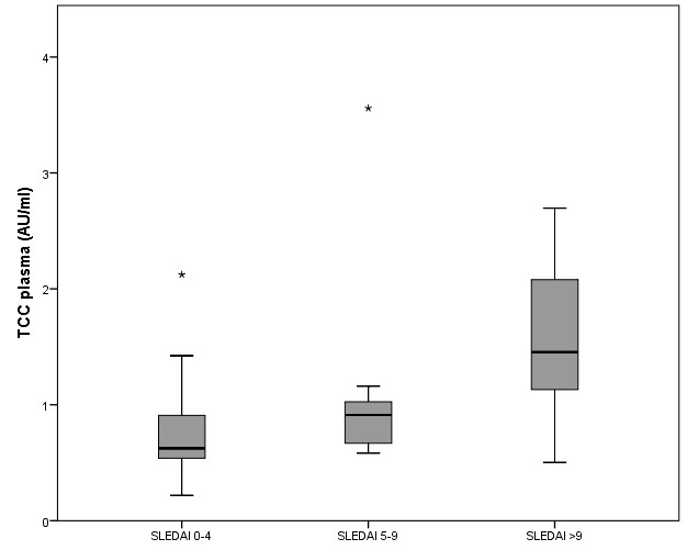
Figure 2: plasma TCC values in the different SLEDAI levels.
Figure 2: plasma TCC values in the different SLEDAI levels.

Figure 3: classical pathway activity in the different SLEDAI levels.

Figure 4: plasma TCC levels as a function of classical pathway activity.
Table 1: TCC concentrations in male and female controls.

Table 2: Median alternative pathway activity in the different SLEDAI levels.

Results
Due to either improper treatment in the preanalytic phase or a lack of sample amount, three plasma samples, six sera, and nine zymosan-activated sera could not be analyzed for the TCC. .sTCC - SLE versus control group The median concentration of TCC in the study group was 0.81 AU/ml in plasma, 7.78 AU/ml in serum, and 658 AU/ml in zymosan-activated serum. There was no statistically significant difference between the concentrations in male and female patients. The median TCC concentrations in the control group were 1.25 AU/ml in plasma, 7.73 AU/ml in serum, and 503 AU/ml in zymosan-activated serum (Tables 1 & 2). Again, there were no significant gender-specific differences.
The Mann-Whitney-U-Test showed no statistically significant differences in the TCC levels between SLE patients and the control group in all three sample types (Figure 1).
sTCC Levels in Correlation to the Disease Activity
Three disease activity categories were defined according to SLEDAI scores. The values of the first group ranged from 0 to 4 (21 samples), those of the second group from 5 to 9 (10 samples), and the third group had SLEDAI scores of 10 or more (8 samples). The Spearman’s rank correlation coefficient measured a significant positive correlation between plasma TCC concentrations and the SLEDAI (p<0.007). (Figure 2)
COMPL 300 – classical pathway
The CP activation mean value in the study group was 56%. According to Seelen et al. [12], the healthy adult population mean value amounts to 98%. The difference is statistically significant (p <0.0001). Twenty-one out of thirty-five (60%) samples were below the cut-off value of 74%. There was also a statistically significant negative correlation between classical pathway activation and the SLEDAI score (p<0.001) as well as the plasma TCC level and the SLEDAI-score (p<0.001) (Figure 3 & 4):
COMPL 300 – alternative pathway
In the AP, 22 out of 35 (63%) SLE patients’ samples were below the cut-off value set at 39 %. The study group’s mean value was 16%. There was no statistically significant difference between male and female patients. The correlation between alternative pathway activity and SLEDAI score was p<0.011.
COMPL 3000 - lectin pathway
Seleen and al. arbitrarily set the lectin pathway cut-off value at 10%. Still, almost one-third of the healthy population's values were below this threshold. Sixty-three percent of the SLE patients’ samples were below 10 %. The median value was 1%. Again, no significant gender-specific difference could be described, but the negative correlation to the SLEDAI was statistically significant (p<0.01).
Anti-CFH-ELISA
All patients in this study tested negative at least twice for CFH antibodies.
Discussion
Regarding sTCC concentrations, both the study group and the controls had the lowest median TCC levels in plasma. The median serum levels were, respectively, six and nine times higher. This phenomenon already occurred in former studies and is most likely due to EDTA in the plasma tubes, inhibiting coagulation and thus in-vitro activation of the complement system [14]. As plasma appears to be the most suitable specimen to reflect in-vivo TCC levels, it was used to conduct the correlations in this study. As expected, the highest concentrations of TCC were measured in zymosan-activated serum, which could reflect the remaining capacity of complement activation in vitro.
Regrettably, we could not provide evidence for a significant difference between median values of TCC in patients with SLE and healthy controls. Therefore, the determination of absolute sTCC levels can not be helpful in the diagnosis of SLE. However, a significant positive correlation between plasma sTCC levels and the corresponding SLEDAI score could be detected in SLE patients. This result indicates that, besides the well-established SLEDAI-Score, the determination of the sTCC is a reliable laboratory marker for the assessment of longitudinal courses of disease in children with SLE. Falk et al. [15] already showed that TCC-concentrations correlate with the disease activity in adults. This study showed that measurements of the sTCC could also be an indicator of disease activity in pediatric patients.
The COMPL 300 showed a decreased functional activation capacity of the CP and AP, while sTCC concentration did not differ between the healthy controls and the study group. This could resemble the relevance of deficiencies in complement activation cascade in the pathogenesis of SLE. Due to low complement activation capacity in the LP in healthy controls, a relevant deficiency of the LP can only be diagnosed with MBL levels measurement in plasma, not conducted in this study. Therefore test results cannot be interpreted correctly. The correlation between SLEDAI-score, complement activation capacity, and sTCC concentration suggests consumption of complement components in acute disease flares. Therefore, the evaluation of the complement pathways via the COMPL 300 is a reliable biomarker for monitoring patients with SLE. With p<0.005, the classical pathway shows the most significant negative correlation and thus confirms that immune complexes cause complement activation in high disease activity.
Approaches to targeting the complement system in systemic lupus have been manifold [16]. Pharmaceutical agents that interfere with complement cascade could be a promising treatment option, especially for otherwise therapy-resistant SLE. In patients with aHUS, where mainly the AP is over-activated, treatment with C5-blockage has shown considerable success [17, 18].
Antibodies against several complement components and their regulatory proteins, like the C1-inhibitor, have been detected in patients with SLE [19]. CFH-antibodies were already found in patients with atypical hemolytic uraemic syndrome (aHUS) – a disease where endothelial damage is also due to permanent uncontrolled complement activation. (13) In our study, no patient tested positive for CFH-antibodies, and consequently, there is no currentindication for a pathogenetic role of this immunoglobulin. This study examined the correlation between disease activity and complement involvement in pediatric patients with SLE. Our findings lead to the conclusion the measurements of sTCC or the complement activation capacity can be valuable additional tools for the assessment of disease activity.
More information regarding this Article visit: OAJBGSR

No comments:
Post a Comment