Tuesday, 16 November 2021

Diagnosis and Treatment of Intramyometrial Placental Tissue Invasion in Cesarean Scar: A Case Report

Diagnosis and Treatment of Intramyometrial Placental Tissue Invasion in Cesarean Scar: A Case Report by Ying Xiao Yan* in Open Access Journal of Biogeneric Science and Research


Abstract

Morbidly adherent placenta is one of the rare complications of Cesarean Section Pregnancy. This is the implantation (part or whole) of the placenta via the niche (dehiscence at the hysterotomy site) or scar of the prior cesarean section into the uterine wall. It is divided into placenta accreta, placenta increta or placenta percreta depending on the degree of placental tissue uterine invasion. It poses a high risk of maternal morbidity and mortality due to missed diagnosis and non-existence of clear treatment guidelines. We present a case of adherent placental tissue in cesarean scar pregnancy diagnosed with laparoscopy. We highlight the importance of early diagnosis and treatment of placental tissue uterine invasion in cesarean scar pregnancy.

Introduction

Cesarean Scar Pregnancy is reported to range from 1 in 1,800 to 1 in 2,500 of all cesarean deliveries performed. Morbidly adherent placenta is one of the rare dangerous complications of cesarean scar pregnancy [3]. Depending on the degree of invasion in the uterine wall, placenta tissue invasion can be classified into accreta, increta and percreta. Over the last few decades, numbers of cesarean section deliveries have increased [1]. This has also led to the increase in the number of uterine scar placental tissue invasion complications like hemorrhage which often leads to many other complications including hypovolemic shock, disseminated intravascular coagulopathy, hysterectomy, and even morbidity. Increase in cesarean section delivery has led to reduced number of vaginal operative procedures [1,2]. ACOG has advocated for vaginal delivery in the trial of scar for appropriate candidates, the rate of repeat cesarean deliveries is now close to 91% [2].

Proposed pathogenesis of cesarean scar pregnancy is that the conceptus invades through a defect or microscopic dehiscence in the scar of previous hysterotomy. This is due to the poor vascularization with fibrosis of the lower uterine segment. There is also disruption of the endometrium and myometrium that predisposes to improper implantation at the site of the prior hysterotomy [3,4]. The placental invasion is due to compromised decidua basalis, which normally is a barrier to trophoblastic invasion of myometrium.

It is important that early and accurate diagnosis is obtained in order to avoid complications. Though missed diagnoses of placental insertion disease (morbidly adherent placenta) have been reported, ultrasonography remains the main tool for diagnosis of morbidly adherent placenta, mostly with a sensitivity of 90.7% [95% confidence interval (CI), 87.2e93.6], specificity 96.9% (95% CI, 96.3e97.5), a positive likelihood ratio of 11 (95% CI, 6e20), and a negative likelihood ratio of 0.16 (95% CI, 0.11e0.23) from a meta-analysis in 2013 [4]. Ultrasonography clues on morbidly adherent placenta include abnormal vasculature, increased size and numbers of vascular sinus, absence of uterovesicle border or retroplacental hypoechoic zone, and invaded placenta insertion on myometrium [3,4].

Case Presentation

We are presenting a female 32-year-old, G3P2, Chinese, Han tribe, initially presented to the local hospital with 3 months amenorrhea. She requested to terminate the pregnancy due to satisfied parity. Cesarean Scar Pregnancy was confirmed at the local hospital with ultrasonography showing 15weeks gestational age. She had no history of bleeding, dizziness, abdominal pain, vaginal discharge, dysuria, diarrhoe, coughing or headache. A known polycystic ovarian syndrome patient, with history of appendectomy and cesarean section delivery 2 years and 8 years ago respectively prior to hospital presentation. According to ultrasound finding, the patient was commenced on medical abortion therapy on the same day of admission, followed by dilatation and curettage on day 2 post admission. During the procedure, patient developed hemorrhage due to retained products of conception (placenta) adherent to the anterior wall of the lower part of the uterine cavity. Estimated blood loss during the procedure was 1500ml. The heavy bleeding in this cesarean scar pregnant patient was treated with uterine artery embolization interventional therapy, blood transfusion and IV fluids. The local hospital decided to refer the patient to our hospital for further evaluation and treatment on the same day she underwent the D&C procedure. Upon arrival in our hospital, patient was seen in the outpatient department with normal vitals and minimal vaginal bleeding noted on vaginal examination. Human chorionic gonadotropin (hCG) was 3562miU/ μL, hemoglobin 81g/L. Hysteroscopy examination revealed retained products of conception tissue in the anterior wall of the lower part of the uterine cavity measuring 5 * 4cm diameter. Under the guidance of B-ultrasound, curettage was done and about 100g of retained products of conception were evacuated. Some of the tissues adhered tightly to the anterior wall of the uterus, and vaginal bleeding was more when scrapped. The procedure was successful and hemostasis was achieved with estimated blood loss of 100ml. Patient was treated with uterotonics to prevent further vaginal bleeding. She was treated as an outpatient to be followed up with a recheck ultrasonography after four days.

One week post curettage, patient was admitted to our hospital due to minor vaginal bleeding mixed with clots. Recheck B-ultrasound examination revealed that the anterior uterus was irregularly enlarged with a heterogeneous hyperechoic mass of 7.3*4.9cm in the middle and lower part of the anterior uterine wall. The mass protruded outward reaching the serosa layer. Color blood flow was seen in and around the mass. There was no obvious abnormality in bilateral adnexa. At this time, placental tissue uterine cesarean scar invasion was suspected.

Discussions with the patient regarding her imaging findings, potential complications of continuation of cesarean scar placental tissue invasion and reproductive goals were done. The patient stated that she desired permanent sterilization. She was scheduled for an urgent laparoscopic removal of the adherent cesarean scar placental tissue and bilateral tubal ligation.

Intraoperatively, during laparoscopic abdominal and pelvic exploration, the uterus showed a subserosal 4*5cm enlarged mass on the lower segment of the uterus as shown is (Figure 1a). The mass was noted to be enlarged with hyper vascularization. Incision on the mass was made to open the intramyometrial mass as observed in (Figure 1b). Access to the intramyometrial space was achieved and complete evacuation of the adherent placental tissue was done (Figure 1c & Figure 1d).

After the evacuation of the intramyometrial adherent placental tissue, the intramyometrial cavity was sutured; uterine scar repair and bilateral tubal ligation were successfully done. Hemostasis was achieved with estimated blood loss of 1000ml. Patient received 4 units of packed red blood cells, 1000ml crystalloid solution and intramyometrial injection of methotrexate. Retained products of conception were taken for pathological assessment which confirmed the presence of degenerated placenta villi and decidua, trophoblastic tissue implanted in an area of markedly attenuated myometrium. Post operatively, patient improved clinically. She had no vaginal bleeding, abdominal pain, dizziness, headache, fever or constipation. Laboratory results showed reduction in hCG levels i.e 558.26 miU/ μL and 234.8 miU/ μL on day 4 and day 7 post operation respectively compared to the hCG on admission of 3562miU/ μL. After 10 days in the hospital, the patient was discharged to be followed up in the gynecology clinic for review.

Figure 1: (a) Laparoscopic view of the pelvic cavity showing the intramyometrial subserosal mass (black arrows). (b) Opening and exploration of the intramyometrial mass review the adherent placenta tissue being evacuated. (c) Evacuated products of conception were sent for pathology examination. (d) View of the intramyometrial cavity after complete evacuation of the products of conception just before suturing it, the intramyometrial cavity did not communicate with the uterine cavity.

Discussion

Morbidly adherent placenta is one of the rare complications of Cesarean Section Pregnancy. This is implantation (part or whole) of the placenta via the niche (dehiscence at the hysterotomy site) or scar of the prior cesarean section into the uterine wall [5,6]. Morbidly adherent placenta is divided into placenta accreta, placenta increta or placenta percreta depending on the degree of placental tissue uterine invasion. Prevalence of Cesarean Scar Pregnancy is reported to range from 1 in 1,800 to 1 in 2,500 of all cesarean deliveries performed [6,7]. Research has shown that, in cesarean section pregnancy, conceptus invades through a defect or microscopic dehiscence in the scar of previous hysterotomy [7]. This is due to the poor vascularization with fibrosis of the lower uterine segment.

Cases of placental tissue cesarean scar invasion are on the rise due to an increase in the number of cesarean sections being performed. Depth of placental invasion increase as the gestation advances. Some of the other risk factors for morbidly adherent placenta include placenta previa after a prior cesarean delivery, a history of uterine surgery (e.g., myomectomy entering the uterine cavity, hysteroscopic removal of intrauterine adhesions, cornual resection of ectopic pregnancy, dilatation and curettage, endometrial ablation), cesarean scar pregnancy, maternal age older than 35 years, history of pelvic irradiation, and infertility and/or infertility procedures (e.g., in vitro fertilization) [8].

Diagnosis of this placental insertion disease is by ultrasonography, mostly with a sensitivity of 90.7% [95% confidence interval (CI), 87.2e93.6], specificity 96.9% (95% CI, 96.3e97.5), a positive likelihood ratio of 11 (95% CI, 6e20), and a negative likelihood ratio of 0.16 (95% CI, 0.11e0.23) from a meta-analysis in 2013 [2,8]. Diagnosis is often difficulty and missed by ultrasonography diagnosis. Magnetic Resonance Imaging has been used as an adjuvant to ultrasound as well as aid in preparation for surgery and intraoperative orientation but it is expensive and has low diagnostic value.

Our case highlights the importance of early diagnosis and management of adherent placental tissue in the cesarean scar to prevent catastrophic patient morbidity or death. If left untreated, patient can develop hemorrhagic shock which may lead to death. No clear treatment guidelines have been suggested for the treatment of Morbidly Adherent Placenta. It has been shown that if cesarean scar pregnancy is complicated with morbidly adherent placenta (cesarean scar adherent placental tissue invasion), the most frequently therapeutic approach is hysterectomy [9].

Conclusion

Our case presentation demonstrates the importance of early diagnosis and treatment of cesarean scar pregnancy, especially if it is complicated with placental tissue uterine scar invasion. The following are highly recommended to prevent serious complications of this condition:

  1. If cesarean scar pregnancy is suspected, dilatation and curettage can be done under the guidance of B-ultrasound or laparoscopy monitoring to avoid puncturing the thin uterine muscle on the scar.
  2. A patient with suspected cesarean scar pregnancy with heavy vaginal bleeding can undergo uterine artery embolization interventional therapy to minimize blood loss.
  3. If a patient does not respond to the 1st blinded dilatation and curettage procedure, a 2nd D&C can be done under the guidance of hysteroscopy monitoring to make sure all products of conception are visualized and evacuated.
  4. For patients with cesarean scar pregnancy and suspected placental tissue implantation, laparoscopy for diagnosis and surgical treatment can be done.

Friday, 12 November 2021

Relation Between Coffee Consumption During Pregnancy and Preeclampsia; A Letter to the Editor

Relation Between Coffee Consumption During Pregnancy and Preeclampsia; A Letter to the Editor by Negin Shaterian* in Open Access Journal of Biogeneric Science and Research


Abstract

Some women continue to drink coffee during pregnancy.  Coffee contains caffeine. There is a concern that caffeine consumption in pregnant women may increase and affect their health and that of the fetus. However, the specific effects of caffeine on the fetus are still unknown [1]. Consumption of less than 300 mg of caffeine during pregnancy, which is equivalent to 3 cups of coffee, does not seem to be a problem [2]. However, some studies have shown that even consumption of less than 300 mg of caffeine leads to Pregnancy failure [2]. Caffeine consumption during pregnancy affects placental-fetal development [1]. In addition, it reduces fetal growth because caffeine easily enters the blood around the placenta and reduces blood flow in the placenta [1].  Therefore, the nutrition and oxygen supply to the fetus is reduced. Low oxygen levels in early pregnancy are an important factor in inducing placental angiogenesis [3]. In the first trimester of pregnancy, when the fetus grows at low oxygen concentrations, it causes proliferation of trophoblast cells [3].  This inflammatory response also occurs before preeclampsia. Therefore, it is possible to confirm the hypothesis that the consumption of caffeinated substances, such as coffee during pregnancy, can lead to preeclampsia. However, a clinical study reported that there was no difference in the meat, fruit, vegetable, coffee and alcohol consumption, smoking, folate use, and oral contraceptives as a possible risk factor of preeclampsia in the current pregnancy [4]. Preeclampsia is one of the serious problems in pregnancy that endangers the health of the mother and fetus.  About 10 to 15% of women develop high blood pressure (140 to 90 and above) after 20 weeks of pregnancy. If this high blood pressure is accompanied by proteinuria, it indicates preeclampsia, which can lead to the development of a serious disorder [5]. About 5,000 women and one million babies die from this disease annually [5]. According to one of the most important indicators in any country is the mortality rate of mothers and infants, so we must identify the factors that lead to an increase in the incidence of preeclampsia and try to eliminate them. However, since the effects of caffeine, even in small amounts, are still unknown, it is best to advise pregnant women not to use caffeinated substances during pregnancy. Moreover, if a person is addicted to caffeinated substances, it is better to be taken care of before pregnancy and after quitting addiction of caffeinated substances, get pregnant. In addition, more clinical studies are recommended to evaluate the effect of caffeinated substances in pregnancy and preeclampsia.

 More information regarding this Article visit: OAJBGSR


Tuesday, 9 November 2021

Expected Life Expectancy of Life Expectants Due to Covid-19 Phenotypes Reduced to Life Expectancy Cohorts

Expected Life Expectancy of Life Expectants Due to Covid-19 Phenotypes Reduced to Life Expectancy Cohorts by Shane Smyth* in Open Access Journal of Biogeneric Science and Research


Opinion

I met a man once who had Covid and he said that he felt it was bad for his health. He had a cough and I said o you poor man, you should go to hospital but he didn’t go to hospital and when I called over to his house two days later to return his Sleepless in Seattle VHS tape, he was dead. I said what are you doing you should have gone to hospital but he did not go to hospital because he was dead and I think he was dead because of the Covid. In his house I found a note that said I have covid and I think I need to go to hospital but I like this scene with Hanks and I should finish it. He also had cooked pasta and beef and I did not eat it but his dog looked hungry and I gave it to his dog but the dog was dead. I don’t know if the dog had covid but I think it’s possible. I then called over to the neighbours and they were alive. I told them that the man and his dog were dead and they asked where was the Seattle tape and I said I thought I might take it home to watch Hanks and they said okay. I said there was some beef and pasta and they said they had sealeeak disease and could not eat it I suppose that’s right. I decided I would give them the Hanks tape and they said even better come inside and we shall watch it. They made tea but when we sat down in the living room, they had a DVD player I see and the VHS did not play. They said what is this 1996 a VHS!? 2021 but they still were laughing that’s okay, I guess. Then I had a dream and what happened was the same. I tell you what the dream was. It happened when I fell asleep on the couch. They had put something in my tea. It was milk. I met a man once who had Covid and he said that he felt it was bad for his health. He had a cough and I said o you poor man, you should go to hospital but he didn’t go to hospital and when I called over to his house two days later to return his Sleepless in Seattle VHS tape, he was dead. I said what are you doing you should have gone to hospital but he did not go to hospital because he was dead and I think he was dead because of the Covid. In his house I found a note that said I have covid and I think I need to go to hospital but I like this scene with Hanks and I should finish it. He also had cooked pasta and beef and I did not eat it but his dog looked hungry and I gave it to his dog but the dog was dead. I don’t know if the dog had covid but I think it’s possible. I then called over to the neighbours and they were alive. I told them that the man and his dog were dead and they asked where was the Seattle tape and I said I thought I might take it home to watch Hanks and they said okay. I said there was some beef and pasta and they said they had seabeach disease and could not eat it I suppose that’s right. I decided I would give them the Hanks tape and they said even better come inside and we shall watch it. They made tea but when we sat down in the living room, they had a DVD player I see and the VHS did not play. They said what is this 1996 a VHS!? 2021 but they still were laughing that’s okay, I guess.

More information regarding this Article visit: OAJBGSR

Friday, 5 November 2021

Effect of Wireless Sensor Networks to Increasing Causes of Numbness

Effect of Wireless Sensor Networks to Increasing Causes of Numbness by Md Rahimullah Miah* in
Open Access Journal of Biogeneric Science and Research


Abstract

Background: Numbness is a complex sensor disease in legs, feet, lower parts of the body or entire body. Yet Medical professionals are facing the insufferable supplementing causes of numbness in individual’s body as a very important global health issue since the 20th century.

Objective: The study aims to evaluate the applications of the fluctuated and processed radio frequencies that effect limbs or other organ of the body within and around the individual’s body boundary at GPS locations. Everyone uses mobile phone within GPS location, but none can know its impact.

Methods: This impact identifies through ISNAH Experiment on cat and dog through application of processed wireless sensor networks at open activities eyes and GPS positions.

Results: This study represents the numbness with sudden pain due to misuse of wireless sensor networks towards an individual’s body at light and dark environment. The research also focuses on the more effective enlarging causes of numbness in dark than light environment. These findings replicate the implication in numbness through operative treatment and recovery that the surgeons provide, which cannot improve efficiently due to abusing wireless sensor networks. The study also found the digitalized health systems are in risks to insecure advances sensor technology.

Conclusion: Systematic healthcare awareness is vital for management with modern technological device but such awareness is still below par, which is alarming to individual’s good health. The study suggests future research trajectories of a new sophisticated alternative secure treatment approach to promote healthcare in the priority of Sustainable Development Goals 2030.

Keywords: Numbness; GPS Location; Body boundary; Sensor Networks; Environment.

Introduction

Nowadays numbness augments in legs or feet or lower part of the body in sudden among human beings [1-4]. Numbness of body is a silent sensor disease, which is numbed from the waist down to the legs [5,6]. Suddenly some male or females may feel numbness in their legs and feet or lower part of the body or entire body because of sitting / sleeping in a position that puts too much pressure on the nerves or reduces blood flow or lack of body electron movement [7-10]. However, continuing or unexplained numbness may be a mark of a causal medical condition [11-13]. Long-term numbness or a tingling feeling in the legs, feet, part of body or entire body may be because of conditions in several ways, such as multiple sclerosis, diabetes, peripheral artery disease, fibromyalgia or unwanted sensor networks [14-18]. A lack of pharmacological tools has delayed our understanding of the physiological mechanisms of causal tingling paresthesia. It may feel the sensation in the entire leg, below the knee, in different areas of foot or lower part of the body within a body boundary of the existing GPS locations [19-22]. From higher study research, the researcher finds new ideas of the reasons any person might experience numbness in the legs, feet or lower part of the body or in the full body suddenly numbness, along with identified symptoms, treatment with recovery systems [17,23-26]. The aim of the study is to find out the root causes of numbness through processed wireless sensor networks to solve with core challenges in worldwide public health security.

Methods and Materials

We conducted this research method at as PhD research work from October 2014 to October 2017 at the Universiti Malaysia Sarawak (UNIMAS), Malaysia. We connected the method with different parameters to enhance data collection, ISNAH Experiment, Specimens Tracking Process, Data Analysis and interpretation as below

Data Collection

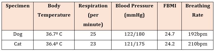
All specimens housed in a room with controlled temperature 36.4°C in cat and 36.7°C in dog with breathing rates, respiration, blood pressure and feline body mass index.The experimental design randomly divided into three experimental groups with Body Mass Index: obese, normal and thin and observed the impact of wireless sensor networks towards pancreases among them in the light and dark environments. The study causes an integration of methods used in wireless sensor networks towards animals’ body and identified its implication. This envisaged the research taking in matter-of-fact research elements to investigate issue hoisted in the study, primarily targeted at SMART devices like telematics’ users towards specimens. Telematics is a smart device, comprises scanner, Global Positioning System (GPS) and Global Navigation Satellite System (GNSS). The fieldwork conducted in the studied area from January 2015 to January 2017.

ISNAH Experiment

Sensor technology comprises ISNAH Experiment. It implies the experiment on the Impact of Sensor Network on Animals and Human beings (ISNAH). The cyber trackers misuse the sensor technology to augment non-communicable diseases among animals and human body. The study examined into two specimens, one is dog and another one is cat among 14 individuals for identification of this misuse application. These animals are available in the study area and suitable for experiment. The study selected sound health two species with Feline Body Mass Index (FBMI) and other following parameters as shown in (Table 1).

The experiment took on dark and light conditions. The specimens stayed in specific geographic location and put the individual inside the iron case (size: 3.5′x 2′x2.5′). Then measurement of individual’s coordinates location includes longitude, latitude and ellipsoid height with GPS and GNSS identifiers.From the field observation, the Automated Radio Telemetry System is more effective in dark than light environment. For this purpose, the study was to examine the system with on smart cell phone, telematics device, iron cage and individual species separately. The dog and cat put into the iron cage with cell phones separately.

The experiment continued at five locations, viz.

  1. Location A with light environments but no Wi-Fi,
  2. Location B with dark and light environments including Wi-Fi,
  3. Location C with dark and light environments including Wi-Fi,
  4. Location D with light environments but no Wi-Fi,
  5. Location E dark and light environments including Wi-Fi.

These experiments continued to identify the reflection of Automated Radio Telemetry System from Telematics device via cell phone towards animals at 09:00 p.m. to 6:00 a.m. from 1 January 2017 to 28 January 2017. The location of experiment settled the species with the temporal conditions through global positioning system. Although many important moments in animal’s life are difficult to study because they are rare, cryptic or occur over large spatial or temporal scales. For the study of FBMI calculation, we used the study web calculator through using rib case circumference and length of the lower back leg from the knee to the ankle.

Table 1: Two selected animals’ specimens with Feline Body Mass Index (FBMI).

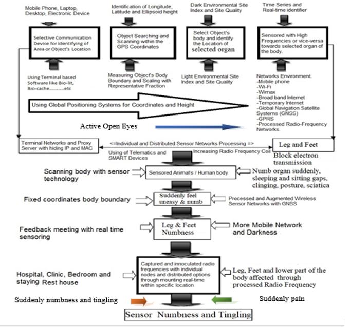
Figure 1: Numbness through Tracking with Processed Radio Frequency [76].

Specimens Tracking Process

Sensor networks track on animals and human beings, where contains in blood circulation and movement of electron. The ISNAH experiment interlinked electron through tracking process. This process included several steps which enhanced to fulfill the Sensored observation. The study was observed the physical conditions including non-communicable diseases of animals like diabetes affected by the telematics device through misapplication radio frequency through tracking process as shown in (Figure 1). Different stages of Tracking Process of Radio Frequency towards animals are listed as below:

  1. Selective communication devices,
  2. Searching object and scanning of individuals body organ,
  3. Identify body organ and light and dark environment,
  4. Sensored the specimens with high, normal and low radio frequency,
  5. Observed and compared the specimens status,
  6. Feedback meeting and illustrated the consequences at result and discussion.

Data Analysis and Interpretation

Quantitative and qualitative related bio-sensor data got through field observation, interviews, field surveys and ISNA experiment while we got secondary data from diverse sources. All general information regarding the occurrence of specimens, status and affected condition checked for accuracy from the unique sources and sources of information also verified. We collected information regarding the initiatives of the authority towards the geographic locations through relevant secondary information and field survey. The compiled and processed data involved in preparing data master sheet and assimilated into suitable systems used in the results and other segments consecutively. The data compiled and analyzed for presentation and interpretation using standard data analysis software like MS Office Suite 2019 and SPSS version 27.

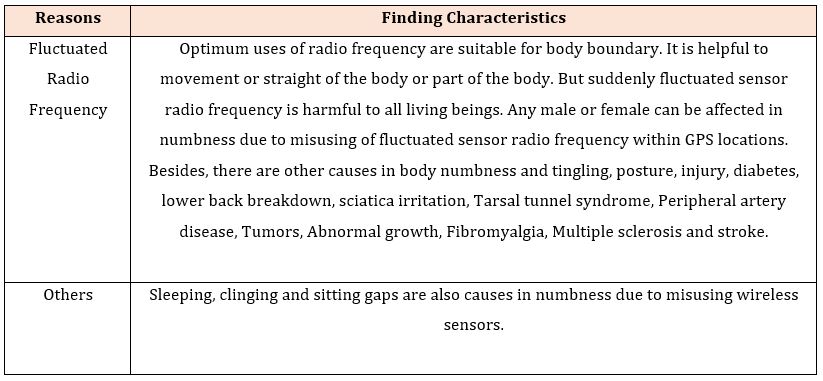
Table 2: Some reasons for numb legs, feet or lower part or whole body due to processed sensors.

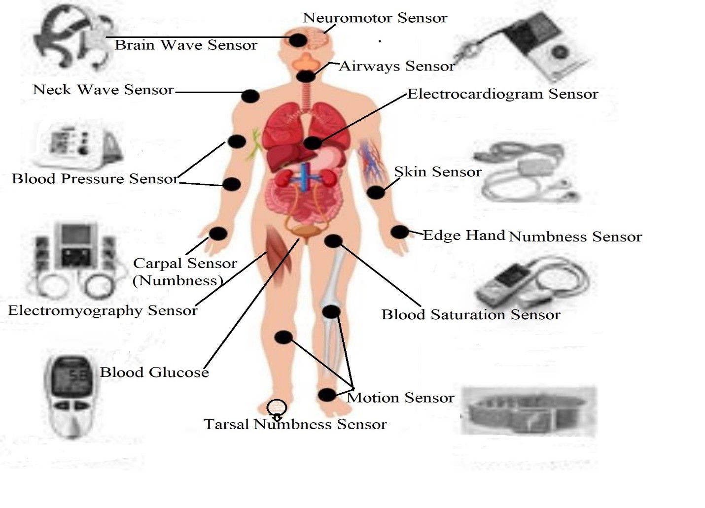
Figure 2: Causes of Numbness of the body or part of the body.

Figure 3: Fluctuated Radio Frequency due to causes of numbness legs.


Figure 3: Table 3: Home-based remedy systems for recovery of legs or feet numb..


Figure 4: Misuse of Processed Wireless Sensor Networks [76].

Results

Occurring of Numbness with Sensor Technology

Due to misuse of fluctuated radio frequencies within a GPS location, any animal or human beings suffers in numbness in actual time within specific radius of the individual’s body boundary. The processed frequencies create the digital poisoning within the circumference of an individual’s optical distance. Because of this, it blocked electron movement or blood circulation with poisoning in GPS location. If any person misuse the telematics or relevant sensor device towards he / she or both of them, they suffer in numbness at the poisoned location.

Suddenly or after a few moments at sitting or sleeping, a male or female’s legs go numb temporarily because of their posture or staying in an existing environment. This numbness creates as a chronic or long-lasting in a specific of the body, which is almost always a sign of an underlying medical condition or physical structure. Suddenly causes of numbness include

  1. fluctuated radio frequency,
  2. posture,
  3. sleeping gaps,
  4. injury,
  5. sitting gap,
  6. diabetes,
  7. clinging gap,
  8. lower back issues,
  9. sciatica,
  10. tarsal tunnel syndrome,
  11. carpal tunnel syndrome,
  12. peripheral artery disease,
  13. tumors,
  14. abnormal growths,
  15. fibromyalgia,
  16. multiple sclerosis,
  17. stroke, and
  18. alcohol use.

There are some specific symptoms for numb legs or feet or lower part of the body. Numbness is just one of the many symptoms associated with temporary and chronic numbness. Many people with numbness in their body organs experience additional symptoms at the same time or intermittingly including

  1. tingling,
  2. burning,
  • tickling,
  1. itching,
  2. crawling feeling under the skin, which as shown in (Figure 2) (Table 2).

If an individual suffers in numbness, he/she can take some simple steps to protect his/her limbs, reducing discomfort and improve mobility. Many people believe it is a crippling and practicable part of growing old. But effects are changing through the expansion of innovative technology. Treatments are better with innovative technology due to open-closed eyes systems, which illustrated in discussion part. Plenty of aged-people are well with little in numbness owing to sensor technological awareness.

The study mentioned that he/she is sitting or sleeping at home or in the office. If he has a mobile phone or sensor device around him with open eyes, his location knew easily. Even if he doesn’t have a mobile phone, we can know your location through laughing, crying, yawning, coughing, sneezing, talking, speaking, flattering, or being with open eyes. The distance between the various parts of individual’s body knew through the telematics device around him, and then digital scanning does with software like Computed Tomographic scan or Magnetic Resonance Imaging, the individual’s hand, legs or lower part of the body or whole body feel numb suddenly. Remote sensing device then applied the fluctuated-frequencies with electromagnetic force to the connective tissue in the area via telematics. After the few moments, fluctuated mobile sensor particles in the blood vessel contracts, the blood flow stops and the connective tissue becomes useless, then the leg or hand or lower part of the body becomes in numbness, which as shown in (Figure 3). In light environment, numbness occurs in 25 minutes and in a dark environment, it takes 12 minutes. However, optimum uses of radio frequency are suitable for body boundary. It is helpful to movement or straight of the body or part of the body. But suddenly fluctuated sensor radio frequency is harmful to all living beings. It can affect any male or female in numbness due to misuse of fluctuated sensor radio frequency within GPS locations.

Discussion

Sensor technology is a blessing to all generations for sound health in a sound mind. But some cyber hackers are misusing the processed radio frequency with sensor technological devices and optical sights within GPS locations [27-30]. They create abnormal situation towards an individual’s body within the body boundary area. Then he/she affects in numbness either temporary or long-lasting [31-34]. Many people say their legs have “fallen asleep”, which is termed in medically as transient paresthesia [35-40]. Habits that can cause the body or part of body to fall asleep including

  1. crossing the legs for too long,
  2. sitting or kneeling for long periods,
  • sitting on the feet,
  1. wearing pants, shoes, socks, gloves that are too tight [41-45].

Most people with fibromyalgia experience a variety of symptoms including

  1. stiffness and soreness for no apparent reason, especially in the morning or after sleeping,
  2. chronic exhaustion,
  3. memory problems and difficulty thinking clearly, sometimes called fibro-fog,
  4. restless leg syndrome [46-50].

Almost everyone with fibromyalgia suffers symptoms in over one part of their body for at least 3 months at a time. Any other symptoms do not accompany if numbness in the legs and feet or is not long term, it is unlikely to be caused by fibromyalgia. The tarsal tunnel is a narrow space on the inside of the ankle [51,52]. Male or female with tarsal tunnel syndrome felt numbness, burning, tingling, and shooting pain in their ankles, heel and feet. It linked this type of nerve damage to reduced levels of B vitamins, such as B-1 (thiamine), B-9 (folate), and B-12, which is caused by excessive alcohol intake [53-56]. The legs are one of the most common parts of the body affected by Peripheral Artery Disease (PAD) [57-59]. Most people with PAD experience pain and cramping in their entire body or part of the body, or in their legs and hips, when they are walking or going upstairs [60-65]. Some are with PAD also experiences leg numbness and weakness. Symptoms of PAD typically go away after a few minutes of rest [66-70]. However, the mentioned causes occurred by the missing of sensor technology, which as shown in (Figure 4). Though they may occur these as usual, either certain conditions. The numbness can cause problems correlated to sensing, feeling, and moving of the body or part of the body [53].

Some patients suffer from the symptoms as numbness in their hands or feet. After lying on one side of the bed at night for a while, the hands and feet on that side feel numb. Because of these reasons, it is difficult to sleep at night. Sometimes, if the patients hold something in their hands for some time, their hands feel numb. After a while they can’t hold on anymore. The doctor observed the patients’ history that they couldn’t even hold the mobile phone in their ears for long while talking on the mobile phones [48]. Many wonders why this could happen, which needs to find the root causes. Because of this problem, the blood circulation in hands and feet is less than normal [71]. Then the pain occurs due to fluctuated pressure on the cervical spine or neck and lumber spine, or the nerves in the waist [56]. The numbness also happens in some diseases including cervical spondylosis, carpal tunnel syndrome, lumber spondylosis, varicose veins, peripheral neuropathy, diabetic neuropathy and motor-neuron disease. Due to lack of vitamins or minerals, the patients suffer in numbness. But the numbness occurs suddenly, the study takes the instant time towards patients’ numbness seriously due to misuse of fluctuated radio frequency. Because of which the numbness later becomes severe, it becomes difficult to get rid of the disease. Therefore, if such symptoms occur, it is necessary to take the advice of a specialist without delay and take treatment for recovery after diagnosing the cause [72]. Occasionally the limbs become numb, which is a symptom of some sensor diseases through misapplications of sensor technology within GPS locations.

Treatment

The proper treatment for numb legs, feet or part of the body depends entirely on the cause.

Medication

Medical options are for long-term numbness in the legs, feet or other organs of the body:

  1. Antidepressants: Some antidepressants, such as duloxetine and milnacipran, have approved to treat fibromyalgia.
  2. Corticosteroids: Some corticosteroids can help reduce chronic inflammation and numbness associated with conditions such as MS.
  3. Gabapentin and pregabalin: Medications that block or change nerve signaling may help reduce numbness associated with conditions such as fibromyalgia, MS, and diabetic neuropathy.

Home-based Remedies

Home remedies that may help to relieve uncomfortable numbness in the legs, feet or lower part of the body through following parameters [73-76], which as shown in (Table 3x).

Challenges

At present, developing countries have no reliable sensor data despite substantial investments in digital health systems due to lack of sensor security [49,50]. The crowdsourcing data about providers, facilities and health measures is likely to grow more individuals with wireless sensor networks in risks. Artificial health intelligence and internet of everything use for better care and diagnosis, but these are expensive innovative technologies.

Conclusion

In conclusion, the study identifies the numbness limbs or part of the body within GPS locations because of the fluctuated radio frequencies. Based on this research, human beings and animals are not secure due to misuse of processed frequencies within body boundary wireless sensor networks in the existing environment. However, the study has attempted to improve a complete scenario of the speeding up causes of numbness in hands, feet, lower part of the body or complete body due to disseminating the fluctuated, homogenous and processed wireless sensor particles. The findings of this study obviously show for sensor network security and dynamic health policy towards present and rationalized generations. Everyone stays at optical sight with body boundary and innovative technology, but none can aware of its security systems. So, policy-makers, health experts and sensor technologists should develop a dynamic, secure health system in the future.

Declarations

Funding

This research work is a part of PhD research, which was funded by the Zamalah Postgraduate Scholarship of Universiti Malaysia Sarawak (UNIMAS), Kota Samarahan, Sarawak, Malaysia and also sponsored by the Information and Communication Technology Division (ICTD), Dhaka, Ministry of Posts, Telecommunication and Information Technology, Government of People’s Republic of Bangladesh. The funders had no role in the design of the research, in data collection, analyses or final interpretation of data, in the writings of the manuscript, or in the decision to publish the findings.

Data Availability

The data are being used to support the findings of this research work are available from the corresponding author upon request.

Competing Interests

The authors declare no potential conflict of interests in this research work.

Acknowledgements

The authors acknowledged the authority of Universiti of Malaysia Sarawak (UNIMAS), Sarawak, Malaysia for providing the Zamalah Postgraduate Scholarship for the completion of the PhD degree. The authors are also grateful to the authority of the Information and Communication Technology Division (ICTD), Ministry of Posts, Telecommunication and Information Technology, Government of People’s Republic of Bangladesh, for a PhD Fellowship during the higher study in Malaysia. The authors acknowledged the Authority of Northeast Medical Pvt. Limited, Sylhet, Bangladesh for kind supports.

Author’s Contributions

MRM designed the study. MRM wrote the first draft of the manuscript with other co-authors ASR, MSK, AAS, MAH, SHC and AKS reviewing and amending the initial draft. AKS was the main supervisor of PhD thesis. All authors read and approved the final version of the manuscript.

More information regarding this Article visit: OAJBGSR

Tuesday, 2 November 2021

Complement Profiling in Childhood-Onset Systemic Lupus Erythematosus

Complement Profiling in Childhood-Onset Systemic Lupus Erythematosus by Hackl L in
Open Access Journal of Biogeneric Science and Research


Abstract

Introduction: Systemic lupus erythematosus (SLE) is an autoimmune disease in which autoantibodies, especially against nuclear components, are the main contributor to its pathogenesis. Complement activation leads to inflammation, and eventually, organ damages occur. Twenty percent of SLE patients are diagnosed in childhood and are known to develop a more aggressive disease course. The etiology of SLE is still subject of the research, but there is considerable evidence attributing its manifestation to genetic susceptibility and environmental factors. This study aimed to investigate the involvement of the complement system in the pathophysiology of childhood-onset SLE. Therefore, the three different activation pathways of the complement system and the soluble terminal complement complex (sTCC) were analyzed. Additionally, we studied the correlation between the systemic lupus erythematosus disease activity index (SLEDAI) and the complement-specific values measured at the time of blood withdrawal. Furthermore, we screened for complement factor H (CFH)-autoantibodies in our study cohort. Finally, the sTCC values were compared to those of an age-matched healthy control group.

Patients and Methods: Blood samples of eight paediatric patients diagnosed with SLE were tested at several points in time for sTCC concentration and activation capacity of the classical (CP), alternative (AP) and lectin pathways (LP). The analytic methods used for all assessments were two different ELISAs.

Results: No significant difference emerged between patients' and controls' absolute sTCC values. However, there was a significant correlation between the patients’ sTCC levels and the SLEDAI score at the time of blood withdrawal (p<0.007). A decreased complement activation capacity was found in 60% of the patients’ CP and 63% of the patients AP and LP. Moreover, the activation capacity of all three pathways showed a significant negative correlation to the SLEDAI score (p<0.001 for CP, p<0.01 for AP, and p<0.01 for LP). No CFH antibodies were detected.

Conclusion: sTCC concentration as well as the extent of activation capacity of the three complement pathways could serve as reliable parameters for monitoring the individual course of disease in paediatric SLE patients. Further research on larger study populations is needed to implement these findings in clinical practice.

Introduction

Systemic lupus erythematosus (SLE) is a chronic autoimmune disease classified as connective tissue disorder. The prevalence ranges from 20 to 70 cases per 100,000 in adults and 3.3 to 8.8 per 100,000 in children [1,2]. Anti-dsDNA antibodies and histones stimulate the circulation of immune complexes leading to inflammation, complement activation, and tissue damage [3]. Deficiencies in the immune regulatory system like decreased phagocytosis of apoptotic material and B-cell hyperactivity are mechanisms underlying the development of the disease [4,5]. Genetic variations that influence lymphocyte activation, immune signaling (e.g., NF-kB, IFNN-I), or clearance of cell debris are associated with SLE susceptibility [5]. The clinical features and disease activity can be variable, with a higher incidence of renal and central nervous system involvement in childhood-onset. [6] The systemic lupus erythematosus disease activity index (SLEDAI) score is commonly used to measure disease activity [7]. As a part of the innate immunity, the complement system and its activation pathways (classical, lectin, and alternative pathways) play a complex role in SLE pathogenesis. Complement deficiencies, especially in the early classical pathway, predispose to the development of SLE, while activation of complement proteins is associated with disease activity [1, 8-10]. Current laboratory diagnostic includes complement protein levels (e.g., C3, C4) and their deposition on cells of the immune system as well as basic inflammatory parameters (e.g., Erythrocyte sedimentation rate, c-reactive-protein) [10].

Methods and Materials

For this observational study, we collected 43 blood samples from eight children (six female and two male) diagnosed with SLE. Sampling took place during clinical consultations at the department of pediatrics and adolescent medicine of the Medical University of Innsbruck between 2014 and 2016. The median age at the time of the procedure was 14, ranging from 4 to 17 years. The samples were tested for the concentration of soluble terminal complement complex (sTCC) as well as for the activation capacity of the three different complement pathways via enzyme-linked immunosorbent assay (ELISA). Results were then compared to those of a healthy control group and put into correlation to the patient’s disease activity at the time of blood withdrawal.

Additionally, all patients were screened for the presence of CFH-antibodies. This study conforms to the Declaration of Helsinki 2000 and was approved by the Ethics Committee. Patients or parents provided informed consent. The collected blood samples were centrifuged, and the pooled plasma or serum was stored in microtubes at -20°C. For each specimen, an aliquot of serum was activated with zymosan (1.5 g baker’s yeast in 10 ml PBS + 0.02 % NaNP).

Prof. Würzner (Institute for Hygiene, Microbiology and Social Medicine of the Medical University ofInnsbruck) established the test protocol for the sTCC-ELISA and generated the anti-C6-, anti-C7- and “WU 13-15”-antibodies. WU 13-15 is targeted against C9 and leads to the fixation of sC5b-9 on the microtiter plate. The second antibody binds either to C6 or to C7 on the complex. Subsequently, alkaline phosphatase-labeled (AP) avidin was used as a second antibody. The substrate for AP is p-nitrophenyl phosphate, and their reaction generates a yellow color [11]. The measuring results of the microplate reader were translated to concentrations with the help of the Microplate Manager Software using a standard curve of maximal activated normal human serum. This standard serum is a mixture of zymosan-activated sera of four healthy individuals, diluted step by step from 1:100 to 1:256,000 in every plate and finally represented a logarithmic calibration curve. As a control group, we used the TCC concentrations of 18 healthy children (10 female and 8 male) between the age of two and 18 years (median: 7 years) that Dr. Thomas Giner already evaluated (unpublished data). All patients came from the department of child and adolescent health of the University Medical Centre of Innsbruck, underwent a routine blood draw, and had no diagnosed inflammation or other complement-related diseases. Their parents signed informed consent for the draw of two additional blood tubes for this study. The samples of the control group were analzsed according to the test protocol of this study.

The WIESLAB© Complement system Screen kit COMPL 300 (Euro Diagnostica, Malmö, Sweden) was used to assess the activation capacity of the three different complement pathways in serum. The implementation was done according to its instruction manual. The microtiter plate was divided into three parts,each coated with an activator of the corresponding pathway. Specific buffers prevented cross-reactions between the different pathways. The complement activation in each pathway leads to the formation of sC5b-9. The concentration of this complex was detected as in the TCC-ELISA described above but expressed as a percentage of the activity of a calibrator serum from healthy individuals contained in the assay kit. The test protocol was based on the work of Seelen et al [12].

The sandwich ELISA kit was used for the quantitative detection of Anti-CFH autoantibodies in the patients' sera. The microtiter plate was coated with human factor H, and avidin-AP-antibodies and the substrate P-nitrophenyl phosphate were added. The final concentration was calculated creating a standard curve out of the extinction values of samples with a known amount of CFH antibodies. The test protocol was based on Dragon-Durey et al. [13].

All the statistical analyses were performed with IBM SPSS Statistics 23 for Windows (IBM Corporation, Armonk, New York, USA). Histograms, boxplots, and the Kolmogorov-Smirnov-Test were used to evaluate the normal distribution of a variable. The Mann-Whitney U-test calculated differences between specific groups,p-values below 0.05 were considered statistically significant. The Spearman’s rank correlation coefficient was used to describe correlations.

Figure 1: plasma and serum TCC levels in SLE patients and healthy control group.

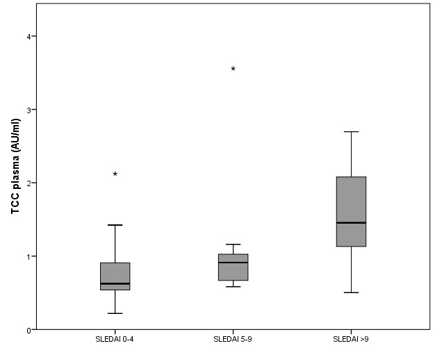
Figure 2: plasma TCC values in the different SLEDAI levels.

Figure 2: plasma TCC values in the different SLEDAI levels.

Figure 3: classical pathway activity in the different SLEDAI levels.

Figure 4: plasma TCC levels as a function of classical pathway activity.

Table 1:  TCC concentrations in male and female controls.

Table 2: Median alternative pathway activity in the different SLEDAI levels.

Results

Due to either improper treatment in the preanalytic phase or a lack of sample amount, three plasma samples, six sera, and nine zymosan-activated sera could not be analyzed for the TCC. .sTCC - SLE versus control group The median concentration of TCC in the study group was 0.81 AU/ml in plasma, 7.78 AU/ml in serum, and 658 AU/ml in zymosan-activated serum. There was no statistically significant difference between the concentrations in male and female patients. The median TCC concentrations in the control group were 1.25 AU/ml in plasma, 7.73 AU/ml in serum, and 503 AU/ml in zymosan-activated serum (Tables 1 & 2). Again, there were no significant gender-specific differences.

The Mann-Whitney-U-Test showed no statistically significant differences in the TCC levels between SLE patients and the control group in all three sample types (Figure 1).

sTCC Levels in Correlation to the Disease Activity

Three disease activity categories were defined according to SLEDAI scores. The values of the first group ranged from 0 to 4 (21 samples), those of the second group from 5 to 9 (10 samples), and the third group had SLEDAI scores of 10 or more (8 samples). The Spearman’s rank correlation coefficient measured a significant positive correlation between plasma TCC concentrations and the SLEDAI (p<0.007). (Figure 2)

COMPL 300 – classical pathway

The CP activation mean value in the study group was 56%. According to Seelen et al. [12], the healthy adult population mean value amounts to 98%. The difference is statistically significant (p <0.0001). Twenty-one out of thirty-five (60%) samples were below the cut-off value of 74%. There was also a statistically significant negative correlation between classical pathway activation and the SLEDAI score (p<0.001) as well as the plasma TCC level and the SLEDAI-score (p<0.001) (Figure 3 & 4):

COMPL 300 – alternative pathway

In the AP, 22 out of 35 (63%) SLE patients’ samples were below the cut-off value set at 39 %. The study group’s mean value was 16%. There was no statistically significant difference between male and female patients. The correlation between alternative pathway activity and SLEDAI score was p<0.011.

COMPL 3000 - lectin pathway

Seleen and al. arbitrarily set the lectin pathway cut-off value at 10%. Still, almost one-third of the healthy population's values were below this threshold. Sixty-three percent of the SLE patients’ samples were below 10 %. The median value was 1%. Again, no significant gender-specific difference could be described, but the negative correlation to the SLEDAI was statistically significant (p<0.01).

Anti-CFH-ELISA

All patients in this study tested negative at least twice for CFH antibodies.

Discussion

Regarding sTCC concentrations, both the study group and the controls had the lowest median TCC levels in plasma. The median serum levels were, respectively, six and nine times higher. This phenomenon already occurred in former studies and is most likely due to EDTA in the plasma tubes, inhibiting coagulation and thus in-vitro activation of the complement system [14]. As plasma appears to be the most suitable specimen to reflect in-vivo TCC levels, it was used to conduct the correlations in this study. As expected, the highest concentrations of TCC were measured in zymosan-activated serum, which could reflect the remaining capacity of complement activation in vitro.

Regrettably, we could not provide evidence for a significant difference between median values of TCC in patients with SLE and healthy controls. Therefore, the determination of absolute sTCC levels can not be helpful in the diagnosis of SLE. However, a significant positive correlation between plasma sTCC levels and the corresponding SLEDAI score could be detected in SLE patients. This result indicates that, besides the well-established SLEDAI-Score, the determination of the sTCC is a reliable laboratory marker for the assessment of longitudinal courses of disease in children with SLE. Falk et al. [15] already showed that TCC-concentrations correlate with the disease activity in adults. This study showed that measurements of the sTCC could also be an indicator of disease activity in pediatric patients.

The COMPL 300 showed a decreased functional activation capacity of the CP and AP, while sTCC concentration did not differ between the healthy controls and the study group. This could resemble the relevance of deficiencies in complement activation cascade in the pathogenesis of SLE. Due to low complement activation capacity in the LP in healthy controls, a relevant deficiency of the LP can only be diagnosed with MBL levels measurement in plasma, not conducted in this study. Therefore test results cannot be interpreted correctly. The correlation between SLEDAI-score, complement activation capacity, and sTCC concentration suggests consumption of complement components in acute disease flares. Therefore, the evaluation of the complement pathways via the COMPL 300 is a reliable biomarker for monitoring patients with SLE. With p<0.005, the classical pathway shows the most significant negative correlation and thus confirms that immune complexes cause complement activation in high disease activity.

Approaches to targeting the complement system in systemic lupus have been manifold [16]. Pharmaceutical agents that interfere with complement cascade could be a promising treatment option, especially for otherwise therapy-resistant SLE. In patients with aHUS, where mainly the AP is over-activated, treatment with C5-blockage has shown considerable success [17, 18].

Antibodies against several complement components and their regulatory proteins, like the C1-inhibitor, have been detected in patients with SLE [19]. CFH-antibodies were already found in patients with atypical hemolytic uraemic syndrome (aHUS) – a disease where endothelial damage is also due to permanent uncontrolled complement activation. (13) In our study, no patient tested positive for CFH-antibodies, and consequently, there is no currentindication for a pathogenetic role of this immunoglobulin. This study examined the correlation between disease activity and complement involvement in pediatric patients with SLE. Our findings lead to the conclusion the measurements of sTCC or the complement activation capacity can be valuable additional tools for the assessment of disease activity.

More information regarding this Article visit: OAJBGSR