Tuesday, 24 November 2020

Plant pathology Articles in JBGSR

Citrus Melanose and Quality Degradation of Fruit by this Disease: A Review by Fazal ur Rehman* in Open Access Journal of Biogeneric Science and Research (JBGSR)

Abstract

Citrus melanose, caused by Diaporthe citri Wolf, is a worldwide fungal disease that is prevalent in many citrus growing areas of Pakistan as well as in the world. Out of two stages of Diaporthe citri Wolf i.e. perfect stage and imperfect stage, the perfect stage causes the citrus melanose disease in many citrus species. Extended rainfall with warm environmental conditions favours the disease initiation and development. This disease result in degradation of fruit quality that results in reduction of marketing and export values of fruit. Proper pruning and use of copper based fungicides are advisable for the treatment of citrus melanose. Plantation of susceptible varieties should be avoided in high rainfall areas. In this article, history of citrus melanose, symptoms of disease, pathogen, epidemiology of disease, quality degradation of fruit and integrated management practices has been discused.


Keywords: Diaporthe citri Wolf; Mud cake Melanose: Teleomorph; Anamorph; Copper-based fungicides

History of Citrus Melanose

The perfect and imperfect stages of Diaporthe citri Wolf cause two different citrus diseases with perfect stage causing citrus Melanose and imperfect stage causing Phomopsis stem-end rot Swingle and Webber firstly described citrus melanose disease near Citra, Floridain 1892 [1]. In 1912, Floyd and Stevens published with evidences that stem-end rot and melanose were caused by the same fungus. At that time they were unable to produce the isolates of fungus from the infected leaves showing the symptoms of citrus melanose on inoculation. The comparison of P.citri and D.citri was made by Fawcetin1932 [2].


In 1940, Ruchle and Kuntz demonstrated that the spores of both P.citri and D.citri had anability to cause the symptoms of citrus melanose [3].Initialy, D.citri was given the name of D.melusaea. But on later, D.citri was given the priority over D.melusaea by Fisher in1972 [3].P.cytosporela was also used for P.citri and it was also proposed that P.cytosporella should be used in place of P.citri. But this proposal was never adopted. The genus Diaporthe/ Phemopsis was place dinphylum Ascomycota with class and order as Sordariomycetes and Diaporthales respectively [4][5]. Four species were found to be different from F.valgure based on their molecular, morphological and cultural data [6]. According to the studies of Gome they have a wide range of host and have ability to form colony on the host that may be diseased plant or dead plant [7]. It was recognized as them most actively transmitting pathogen of citrus.

Symptoms of the Disease

The citrus melanose disease, which is superficial and has no effects on internal quality of fruit but cause the external quality degradation, produces its symptoms on fruits, leaves and small twigs. In case of foliage, the symptoms appear with the formation of small water-soaked speaks that on later become centrally depressed and surrounded by undepressed translucent yellow arcas [8]. After a week, the exudation of gummy substances occurs due to the rapture of leaf cuticle that on later become brown in colour and hardened. These areas have sandpaper- like texture. In case of severe infection, the leaves become pale green to yelow.


The infections may occur on the green twigs .In case of severe infection, the defoliation Occurs that induces the twigs dieback [9].On fruit, scattered speaks are formed in case of high infestation. The infection on young fruits may causet the premature abscission of fruit. In case of late infection, flater pustules are produced. In severe cases, firstly the formation of solid patches of blemish and then cracking of fruit surface occurs, this condition is called mud cake melanose [10]. On the fruit surface, there is also formation of tear-streaks and water droplets patern. The formation of star melanose occurs by the late application of copper based fungicides on the diseased portion of plant because of formation of dark and corky lesions, more prominent then normal and are in star shape [11].

Pictorial Description of Symptoms: (Figures 1 & 2)
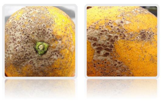
Figure1: Symptomatic lesions of Citrus Melanose present on fruit surface.
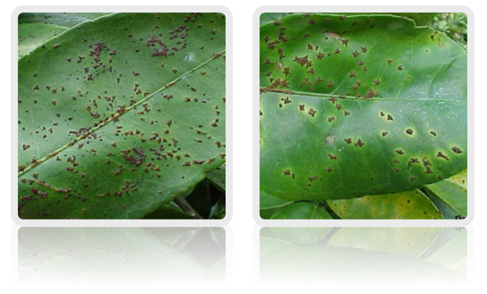
Figure 2: Lesions on leaf surface and formation of Star Melanose symptom on leaf.

Pathogen

The Teleomorph of fungal pathogen is Diaporthe citri Wolf and anamorph is Phomopsis citri Fawc. Mostly leaves and fruits are infected by Diaporthe citri Wolfand Phemopsis citri attacks on stem and causes the stem-end rot [11]. In citrus, fungal pathogen also acts as saprophyte for the infestation on dead twigs. The hyline ascospores of Diaporthe citri produced in each cell are slightly constructed at septum and are in the form of oil droplets. Ascospores are produced in flask shape perithecium. The average size of ascospores is 12.85 microns by 3.85 microns with the perithecium size on average is 500 microns by 50 microns at beak and the base is of diameter125to160microns[12].


The perithecium are projected outward from the stem. The ascospores are ejected from the perithecium forcefully and then they become air borne and spread out over the large distance [1]. On culture media, fan-shaped, white colored mycelia are produced [13]. When spores get substrates, they produce septate hyphae [12]. In disease cycle, the most important state of fungal pathogen is its conidial state. Two types of conidia i.e. alpha conidia and beta conidia are produced by the pathogen. The primary source of spread of fungal pathogen and with the size range from 5-9 microns by 2.5-4 microns are alpha conidia tha are hyaline and single celled. The beta conidia that are slender-rod shaped and hook-like at the end, are spread with great efficiency during rainy condition over nearby substrate and from mycelium [12].

Epidemiology of Disease

The attack of fungal pathogen causing citrus melanose is during the immature stage of foliage, fruits and twigs. Because when the tissues are matured, they mostly become more resistant to the attack of pathogen. Therefore, the susceptibility period for the attack of pathogen over the plant is the period of first 8 to9 weeks after their formation. The formation of symptoms of melanose can vary according to the level and the time of infection. The flyspeck melanose symptoms are produced at the end stage of susceptibility period [14].


As the discharge of ascospores is forcefully and they can be dispersed over the long distance, therefore, the inoculums of fungal pathogen can be spread over the large distance. That's why, there is an increase in the cases of infection be cause of wide spread of large number of ascospores [15]. The disease wil be initiated when the ascospores or conidia of Diaporthe stage or Phemopsis stage land on the surface of plant tisue. The favourable environmental conditions for the infection include dry conditions and the temperature ranging between 17 to 35°C. Above or below this temperature the spores mostly die and the chances of infection reduced [16].


About 10 to 24 hours of moisture are required for the germination of spores but approximately 36 to 48 hours are required for the germination as well as the formation of germ tube that directly penetrates into the tissue of cuticle layer [16]. Then the infection of citrus Melanose pathogen starts.

Quality Degradation of Fruit

Melanose is a serious disease of citrus plant which results in small spots on the surface of citrus fruit. These spots increase in number as the disease reaches its serious stage. These spots cover the entire surface of fruit and reduce the aesthetic quality of fruit. When the disease affects the young fruit, they remain small in size and fall of before reaching maturation stage. In this way, it also results in quality reduction. Atadvanced stage of disease, the spots produced are more solid and the surface of fruit becomes cracked. The fruit affected by melanose disease is not preferable for marketing and export purpose due to quality degradation and thus the market value and export of citrus reduced because the quality of citrus fruit is reduced. However, the disease does not affect the pulp so the fruit for processing purpose is not generally affected but the marketing and export of the diseased fruit is reduced due to the quality degradation and this may cause serious economic losses.


Melanose is a severe disease of citrus in most citrus producing countries and mostly the grape fruit and lemon are affected by this disease [17]. The quality and value of disease afected fruit is reduced and it is not accepted by consumers as a fresh fruit and marketing value is also reduced. The diseased fruit is used as low grade fruit for processing purposes. Farmers and exporters are economically affected due to value loss of citrus fruit by melanose disease.

Integrated management practices

Although, there is no any impact of disease over the yield, so the juice and processing also Remains unaffected But the quality of the fruit for marketing and export purpose is adversely afected. So following integrated management practices should be done for the control of quality losses and fruit degradation by citrus melanose.
a.        Dead branches should be prune out periodicaly. The pruning will help to increase the circulation of air through canopy of plant to keep it dry and the sites for the survival of saprophytic pathogen will also be reduced. It will also enhance the effective penetration of fungicides through for foliage [18].
b.       The fungicides should also be applied for the the disease control. Mostly the application of Copper-based fungicides is done worldwide. The symptoms of star melanose can also be produced over the application of Copper-based fungicides that are not the actual symptoms of this disease [19].
c.        The plantation of susceptible varieties including sweet orange, grape fruit and pomelo should be avoided in high rainfall areas [20].
d.       Other management practices should be done including plantation of citrus in low rainfall and sunny areas, proper sanitation should be done, and intercropping should be avoided.

Conclusion

The symptoms, signs, signals of fever are only seen at the presence of fever. During cancer, the symptom, signs, and signals of cancer are shown every time. A patient having cancer and fever at the same time, symptoms, signs, and signals of both cancer and fever are shown every time. A symptom of cancer never becomes a symptom of fever or a symptom of fever can never become a symptom of cancer.


If fever is a symptom, one symptom has no ability to make other symptoms

 Fever makes numerous symptoms, signals, and actions, etc.


If fever is a symptom we cannot call a person as a fever patient


If fever is a symptom no treatment is required to reduce fever. Treatment is required only for disease and its cause. Our immune system never increase elevated the symptom in the hypothalamus.


Tuesday, 17 November 2020

Dentistry Articles in JBGSR

Higligting the Role of Human Oral Microbiome and Theire Relationship among Most Prevelent Human Oral Cavity Diseases Caused by Bacteria by Bayan Mohammed Fagera* in Open Access Journal of Biogeneric Science and Research (OAJBGSR)

Introduction

The human oral cavity includes numbers of different habitats, such as teeth, tongue, cheeks, and hard and soft palates, and it is harbored by thousands of bacterial species [1]. The human microorganisms found in human oral cavity have been represented as the oral microflora, oral microbiota, or more lately as the oral microbiome. This term microbiome was presented by Joshua Lederberg who is an American geneticist and pioneer in the field of Bacterial genetics also he is one of the researchers involved in The NIH (Human Microbiome Project) [2]. The oral microbiome is extremely multifarious. The average adult harboring about fifty to one hundred billion bacteria in the human oral cavity, which characterize about two hundred prevalent bacterial species. There are nearly about seven hundred prevalent taxa of which less than one third have not yet been cultured in vitro [3]. The oral cavity is the main entrance to the human body. The majority of oral microorganisms are commensal which plays a very significant role in sustaining the steadiness of the mouth ecosystem. In spite of this, some of these microorganisms shows an important role in some oral disease specifically dental caries and periodontitis as shown in Figure 1[4].


Figure 1: Model depicting host-microbe interactions in the pathogenesis of dental caries. 


Each human body is composed of a specified microbiome that is crucial to maintain a good health but also able to elicit disease. The oral microbiome is especially imperative to health because it can cause both oral and systemic disease. The oral microbiome rests within biofilms throughout the mouth, forming an ecosystem that maintains health when in equilibrium. However, certain ecological shifts within the microbiome allow pathogens to manifest and cause disease. Severe sorts of oral disease may end in systemic disease at different body sites. Unlike most infectious diseases where a one causing agent are often found in charge for the infection, oral diseases appear to be the result of multiple microorganisms. In periodontitis, as an example, minimum of three bacterial organisms are found to be directly related with the occurrence of the disease. The mouth harbors an abundant and diverse microflora, which is usually found within biofilms attached to the varied soft- and hard-tissue surfaces. Recent studies using molecular methods have revealed previously unrecognized species within biofilms associated with health and several common oral diseases. These unrecognized species established an under-appreciated diversity within the flora, with new inquiries to be answered. Information regarding the composition of the oral microbiome associated with oral health, dental caries, periodontal disease [5].

Oral Microbiome Composition

The human oral cavity is composed of various microorganisms such as Bacteria, Viruses, Fungi and Archaea [6].

Bacteria

Bacteria account for the most portion of oral microorganisms, and the major knowledge of the composition of oral bacteria comes from past culture-dependent methods. Culture-dependent techniques led to the identification of specific microorganisms thought to possess a causative role in caries and periodontitis [7]. The oral bacterial community is dominated by the 6 major phyla, Bacteroidetes, Proteobacteria, Actinobacteria, Firmicutes, Fusobacteria and Spirochetes, which account for ninety four percent of the taxa detected. (http://www.homd.org).

Viruses

Most of the viruses in the mouth are related to diseases. Such as Herpes simplex Virus which causes primary herpetic gingivostomatitis (is the most common specific clinical appearance of primary herpes simplex infection in childhood) [8]. and recurrent lesions on the face and lips [9] another common mouth viruses is Epstein-Barr virus (EBV) And cytomegaloviruses(CMV)  which are presented in a large majority of adults, but in utmost cases most likely without ever causing any obvious disease. Both viruses however can cause mononucleosis; EBV being in charge for most of the cases. Mononucleosis is known as ‘‘kissing disease’’ signifying that the virus spread through the direct contact mouth-to-mouth [10].

Archaea

Archaea constitutes just a slight part of the oral microbiome and it is restricted to partial species. The founded species were Methanosarcina, Methanobrevibacter, Methanobacterium, Methanosphaera and Thermoplasmatales which all are methanogens [9,11].

Fungi

Fungi are presented extensively in the oral cavity. Not just as an opportunistic pathogen of the elderly and immune-compromised people, but also as part of the mouth ecosystem. The fungal species in the human oral cavity of each individual ranges between nine and twenty-three. The Candida species were the most common, pursued by Cladosporium, Aureobasidium, Saccharomyces, Aspergillus, Fusarium, and Cryptococcus [12].

Location of the Oral Microbiome

The oral ecosystem is extremely intricate because it has quite a few significantly different niches, including saliva, soft tissue surfaces of the oral mucosa and tongue, and hard tissue surfaces of teeth [13]. Since The tissue surfaces and biofilms of the oral cavity are continuously infiltrated with saliva, The salivary microorganisms mainly come from the shedding of the biofilm on the surface of the oral tissue [14] Therefore, the microbial profile of saliva is analogous there to that of the soft tissues, but saliva and soft tissue colonization differ obviously from that of dental plaque [15].

Oral Health

Relative amounts of the microbiome are influenced by factors related   with modern daily life, such as general dieting patterns, sugar consumption, tobacco smoking, oral sanitization, consuming  of antibiotics and other antimicrobials [16]. It is clear that the biofilms related with healthy teeth and gums is portrayed by a very limited commensal microflora ruled by the phylum Firmicutes, as well as a diverse group of streptococcal species. These microorganisms certainly have benefits to the host by interacting with the colonization of exogenous pathogens. This occurs through the inhibition of adhesion of pathogens by commensals or by the production of toxic products such as (bacteriocins, hydrogen peroxide, etc.) to inhibit the growth of pathogens [17].

Oral Disease

Since The oral microorganisms plays a significant role in the health condition of the host and it is an important component in a various oral and non-oral diseases. such as Dental Caries, Periodontal Diseases, and Endodontic Infections.

Dental caries

Dental caries is one of the most frequent chronic infectious diseases worldwide and threatens humans throughout their life, not only during childhood or adolescence. It is also the most common cause of tooth loss and pain in the oral cavity [18,19].it is defined as the physical and chemical processes of demineralization and remineralization occurring on the   surface of the tooth  [20].

Periodontal diseases

Periodontal diseases are acute, infectious, inflammatory diseases that have a multifactorial character in terms of etiology and pathogenesis. Periodontal diseases are divided into two major categories as gingivitis and periodontitis both of which are chronic infectious and inflammatory diseases. Microorganisms and/or their virulence factors trigger the host response and the activated immune cells cause periodontal tissue destruction [21].

Endodontic Infections

in its healthy and intact state, the root canal system is free of infections. Unlike the mouth, the basis canal system has no commensal microbiota, and any microorganism detected here are often considered a possible pathogen. Once microorganisms find their way into the basis canal system, the results may vary from an easy reversible pulpitis to the necrosis of   pulpal tissue and in the end to formation of a periapical lesion: apical periodontitis. Pulpal necrosis on its own, when no microorganisms are involved, doesn't necessarily cause apical periodontitis [22,23].

Conclusion

Based on various researches oral microbiome is proven to be very crucial aspect in oral diseases and general health of humans, keeping a good oral hygiene and good diet is an important thing to prevent some serious oral disease. More research on the oral microbiome should be done to get the overall picture of the whole microbial community inhabiting the human oral cavity.  


Monday, 9 November 2020

Anesthesia Articles in JBGSR

Anesthesia Management in Citoreductive Surgery and Hypertermic Intraperitoneal Chemotherapy Cases by Ugur Koca in Open Access journal of Biogeneric Science and Research (OAJBGSR)

Abstract

Hyperthermic intraperitoneal chemotherapy (HIPEC) applied in conjunction with cytoreductive surgery (SRC) is an effective multimodal treatment option that has been applied in recent years, especially in selected cases of peritoneal malignancies such as peritoneal carcinomatosis, pseudomixoma peritonei and primary peritoneal tumors [1,2]. Cytoreductive Surgery involves the excision of macroscopic tumors and visceral and / or parietal peritonectomy in a single session or a series of operations, ranging from isolated omentectomy to removal of the gastrointestinal tract, pancreas, spleen, bladder, uterus, ovaries and liver. The purpose is to clean all tumor tissue up to 2.5 mm, and to ensure that the rest is affected with a cytotoxic agent. The success of cytoreductive surgery and the prediction of 5-year survival depend on the peritoneal cancer index [3] and abdominal eksplorasyon depends on the time it was made [4-7].


In hyperthermic intraperitoneal chemotherapy, the purpose is to eliminate the tumor tissue at microscopic level by applying chemotherapeutic perfusate prepared to all quadrants at 41-42 °C via a special pump.This major surgery and HIPEC application, which has high morbidity (25-41%) and mortality (0-8%), is not only for surgeons and oncologists, but also for anesthesiologists. Conditions of interest to an anesthesiologist are the purpose and objectives of the operation, the anticipated metabolic and physiological disturbances, and sthe possible chemotherapeutic toxicity [7]. For this reason, between surgeon and anesthesiologist’s cooperation and information sharing is very important. The team must be alert to the cardiovascular status, oxygen consumption, hypo and, or hyperthermia, pain management, and coagulation status in these patients and must be able to make collective decisions in perioperative management.

Hyperthermic Intraperitoneal Chemotherapy

It is the maximum exposure of tissues exposed to chemotherapeutic agents at doses 20-1000 times higher than the targeted plasma levels during hyperthermic intraperitoneal chemotherapy procedure and minimum exposure of normal tissue. HIPEC drugs, they are high molecular weight hydrophilic agents that cannot cross the peritoneal fluid-plasma barrier and their peritoneal clearance is slow. It shows its effect by creating a direct cytotoxic effect and immune-mediated attack in tumor cells through hyperthermia, inhibition of DNA repair mechanisms, protein denaturation and activation of heat shock proteins. Hyperthermic intraperitoneal chemotherapy is more effective when applied immediately after SRC. When this procedure is applied before the gastrointestinal tract reconstruction, it prevents the maling cells from settling into the scar tissue, adhesion and anastomosis sites. HIPEC can be applied with a closed or open abdominal technique (the abdomen remains open during the procedure) Advantages of the closed technique: Reduced heat loss, Increased tissue penetration with the effect of increased intra-abdominal pressure and Reduced contamination risk [8,9].


In the intraoperative period in HIPEC application with the closed abdomen technique, following peritonectomy procedures, it is performed in the abdomen with one / two suprahepatic inflow and two / three pelvic outflow catheters. Perfusate circulates in the abdominal cavity using a Roller pump at 42-43 °C [10]. The temperature is monitored throughout the procedure with multiple probes placed in different areas within the peritoneal cavity. Heated cytotoxic agents are added to the perfusate and HIPEC application takes 30-90 minutes according to different protocols. Following the perfusion of the chemotherapeutic agent, abdominal lavage, drainage and closure of the abdomen are performed. Compared to early postoperative intraperitoneal chemotherapy, it is more effective on survival time. The results are better than normotermic intraperitoneal chemotherapy, and compared to systemic chemotherapy, the average survival time is prolonged by 16-24 months and the 5-year survival rate is increased by 30-45% [11].


 During this procedure, the toxic effects of chemotherapeutics, which type of carrier solution is used and how much is important for an anesthesiologist. Although isotonic solutions or dextrose-based peritoneal fluids are generally used, 5% dextrose-based water solutions are used, as chloride ions for Oxaliplatin alone will reduce Oxaliplatin to less cytotoxic metabolites. This can cause hyperglycemia, metabolic acidosis, and hyponatraemia [11,12]. There is also an increased risk of intraperitoneal bleeding and thrombocytopenia for hypertonic carrier solutions [12-14]. In addition, due to the use of Cisplatinium (half-life 20-30 minutes), as a result of renal loss of Mg, prolongation of QT (pre and intraoperative Mg levels are important) and deterioration in hemodynamic functions with direct cardiotoxic effect may occur [14,15]. 

Physiopathological Changes During Cytoreductive Surgery and Hipec

During this aggressive treatment, many physiopathological changes occur in vital function and parameters within hours. These initially develop secondary to major surgery, and eventually due to hyperthermia and increased intraabdominal pressure. Hypothermia due to excessive fluid loss in the cytoreductive phase. During hyperthermic intraperitoneal chemotherapy, intraperitoneal application of hot solutions increases body temperature up to 40.5°C (mean 37.5 °C). Increased body temperature increases the metabolic rate: increased heart rate, end tidal CO2 level, metabolic acidosis and arterial lactate levels increase in systemic oxygen demand, which reaches maximum levels at the end of the HIPEC period. In the cytoreductive phase, excessive fluid loss may occur due to acid drainage. Afterwards, filling the abdomen with perfusate during the closed HIPEC period causes an increase in intra-abdominal pressure. This causes the diaphragm to shift towards the cranial, leading to a decrease in functional residual capacity and an increase in airway pressure.

These changes cause a sudden increase in central venous pressure by affecting the decrease in oxygenation and cardiac output.


This is also associated with decreased abdominal blood volume and increased splenic vascular resistance. Cardiac output and heart rate can be measured as high due to the hyperthermic intraperitoneal solution used during hyperthermic intraperitoneal chemotherapy and the increased metabolic rate. The initial response to heat stress is peripheral vascular dilatation, which increases heat loss from the center to the periphery. Heart rate increases in order to maintain the increased cardiac output due to decreased peripheral resistance. Due to increased intraabdominal pressure, central venous pressure (CVP) is considered to be a poor indicator of reflecting the volume status. Cardiac output measurement may be required with a swan-Ganz catheter and thermodilution method, transesephageal echocardiography or Picco device. In half of the patients intraoperative, and one third of them postoperatively, there is blood loss that requires replacement. Large fluid shifts and protein loss due to excessive fluid turnover can cause coagulation disorders. Fibrinogen, INR and AT III levels decrease, prolongation of aAPTZ level and thrombocytopenia may be seen.

Preoperative Preparation

What is important in the preoperative anesthesia care of these patients is that the systemic absorption of peritoneal fluid caused by this type of operation, blood loss, aAcute kidney damage, aAcid presence / evacuation, eElectrolyte imbalances, hHypothermia and hHyperthermia, as well as surgery and anesthesia maintenance can be difficult. It is to be borne in mind that it can cause failure. Cardiac risk in this group of patients is the same as in other patient groups. What is important is whether these patients can compensate for the operation-specific physiological changes, such as tachycardia, increased cardiac index and increased oxygen consumption. In patients with advanced age or risk in cardiac tests, the American College of Cardiology / American Heart Association Noncardiac Surgery Quideline should be followed [15,16]. Preoperative mandatory laboratory examination consists of electrolytes, blood urea nitrogen and creatinine level, albumin, bilirubin, hemogram, coagulation levels and glucose levels.


Although the renal damage associated with HIPEC is reversible, the level of the preoperative calculated glomerular filtration rate in these cases is also accepted as a postoperative renal damage indicator. Also, the presence of preoperative renal dysfunction was found to be associated with perioperative cardiovascular events [16,17].

Conditions to be Considered In Intraoperative Anesthesia Management

In the cytoreductive phase, hypothermia develops due to excessive fluid loss. One of the most important issues in the intraoperative anesthesia management of these patients is the risk of hyperthermia that may occur by bringing the carrier solution to 42-43 °C (mean 37.7 °C, sometimes 40.5 °C). Hyperthermia; Iit can cause coagulopathy, arrhythmia, liver / kidney damage, peripheral neuropathy and convulsions. Therefore, controlled hypothermia application before HIPEC may be preferred by methods such as reducing the operating room temperature, application of cooled intravenous fluid or not using a surface warmer. However, although the risk of hypothermia is more tolerable against hyperthermia, it should be kept in mind that hypothermia may lead to changes in anesthetic drug pharmacokinetics and increase in blood loss, the risk of surgical site infection and adverse cardiovascular effects [17-21].


With total body hyperthermia, heart rate, cardiac index and oxygen consumption increase, systemic vascular resistance decreases. Plasma norepinephrine levels increase in parallel with the increase in temperature. Therefore, radial artery catheter and invasive arterial monitoring should be added to the standard monitoring recommended by ASA in these patients. Central venous pressure monitoring due to increased intraabdominal pressure is considered a poor indicator in reflecting the volume status. Cardiac output measurement may be required with a swan-Ganz catheter and thermodilution method, transesephageal echocardiography or Picco device [19-23].


In the cytoreductive phase, excessive fluid loss may occur due to acid drainage. Afterwards, filling the abdomen with perfusate during the closed HIPEC period causes an increase in intra-abdominal pressure. This causes the diaphragm to shift cranially, resulting in a decrease in functional residual capacity and an increase in peak airway pressure. As a result, deterioration in oxygenation, sudden increase in central venous pressure, Vena cava inferior compression, decrease in abdominal blood volume, increase in splenic vascular resistance, decrease in preload and increase in gastric pCO2 and pH decrease (microcirculation effect) are observed. Intraabdonminmal pressure can reach up to 12-26 mmHg and this requires good muscle relaxation [24].


These changes cause a decrease in oxygenation and a sudden increase in central venous pressure by affecting cardiac output. This is also associated with decreased abdominal blood volume and increased splenic vascular resistance. In half of the patients intraoperative, and one third of them postoperatively, there is blood loss that requires replacement. Bleeding is not only associated with surgical reasons, but also with large fluid shifts, hyperthermic chemotherapy and protein loss due to excessive fluid turnover. There is a decrease in Fibrinogen and AT III level, prolongation in Fibrinogen, INR and aPTT, and a decrease in coagulation factors such as Thrombocytopenia and F XIII [25-27].


In these cases, blood volume plays a major role in the maintenance of systemic and regional perfusion in the intraoperative period. Decreased regional perfusion is a cause of acute renal failure. For the prevention of acute renal failure, administration of Ffurosemide (mean 25 mg), diuresis control and normovolemia are important. Acid drainage and excessive debulking can cause protein loss of up to 700 g / dayG. Therefore, in some centers, coagulopathy and accompanying albumin deficiency - if albumin <1.5-2 g - are also performed with fresh frozen plasma and albumin replacement [28-29].

Features of the Postoperative Period

In these cases, follow-up in the intensive care or recovery unit may be considered due to multiple organ failure that may develop due to disorders in the perioperative period or physiological changes. The important thing is to monitor the organ functions with constant close monitoring and to start continous positive airway pressure (CPAP) application in order to provide adequate oxygenation in these cases when necessary. In this period, management of intraoperative complications and, if any, coagulopathy and / or metabolic disorders should be corrected in the early period. In order to maintain pneumatic compression initiated in the operating room for deep vein thrombosis prophylaxis until mobilization and to prevent postoperative ileus, it is important to start oral intake early and to provide postoperative pain with epidural analgesia, if possible [30-32].


Expected risks specific to the postoperative period; Intestinal perforation, Anastomotic leak, Bile leak, Fistula, Pancreatitis, Postoperative bleeding, Deep vein thrombosis, Pulmonary embolism and wound opening. Also, Volume expanders and / or Norepinephrine or vasopressin may be used depending on the post-HIPEC vasodilation (SIRS). However, regardless of fluid response, aggressive fluid loading It may cause an increase in cardiac filling pressures and pulmonary edema [33].